People pay deerly for the switch from daylight saving time.
The change to standard time in autumn corresponds with an average 16 percent increase in deer-vehicle collisions in the United States, scientists report November 2 in Current Biology. The researchers estimate that eliminating the switch could save nearly 37,000 deer — and 33 human lives.
In a typical year, there are more than 2 million deer-vehicle collisions — about 7 percent of total vehicle crashes. To see how much the biannual time change impacts those numbers, wildlife biologist Laura Prugh and colleagues compiled data from 23 states that tracked whether a crash involved an animal and what time the crash occurred. The team compared those numbers to traffic volumes for each state between 2013 and 2019, focusing on the weeks before and after the switches to daylight saving time in springtime and back to standard time come fall.
Springing forward had little effect, but almost 10 percent of yearly deer collisions on average took place around the autumn fallback — when the bulk of human traffic shifted to after dark. The problem was especially acute on the East Coast. “You see [a] really steep spike in the fall,” says Prugh, of the University of Washington in Seattle. “In the western states, you also see an increase, but it’s not nearly as sharp.” On the East Coast, the autumn switch falls in the middle of mating season for white-tailed deer. Not only are more drivers active after dark, more deer are too. “The timing could not be worse.”
Eliminating the clock change wouldn’t completely wipe out the spike in crashes — mating season plays a big role, regardless of what time sunset happens. But the scientists estimate that keeping daylight saving time year-round would decrease total deer-human collisions by about 2 percent — saving dozens of people, thousands of human injuries and tens of thousands of deer. It’s another reason for us all to move toward the light (SN: 3/31/14).
The monument consisted of a circle of immense, finely tooled stone archways surrounded by a range of 56 equally spaced [holes].… The precisely proportioned placement of the stones and holes has led archaeologists to presume that the monument had some great astrological significance.… As an alternate explanation, the researchers say perhaps there were 56 families, clans or social units who built Stonehenge and who were entitled to dig one of the [holes] and use it to inter cremated remains.
Update Stonehenge’s purpose remains murky, but the monument’s origin is becoming clearer thanks to science. For at least the first 500 years of its existence, Stonehenge was a cemetery (SN: 5/29/08). A chemical analysis of remains at the site suggests that some of the people interred there came from Wales, more than 200 kilometers west of where Stonehenge stands in southern England (SN: 8/2/18). The monument’s first building blocks also may have come from Wales, repurposed from a stone circle there, but that hypothesis is debated (SN: 2/11/21).
An ancient, armored worm may be the key to unraveling the evolutionary history of a diverse collection of marine invertebrates.
Discovered in China, a roughly 520-million-year-old fossil of the newly identified worm, dubbed Wufengella, might be the missing link between three of the phyla that constitute a cadre of sea creatures called lophophorates.
Based on a genetic analysis, Wufengella is probably the common ancestor that connects brachiopods, bryozoans and phoronid worms, paleontologist Jakob Vinther and colleagues report September 27 in Current Biology.
“We had been speculating that [the common ancestor] may have been some wormy animal that had plates on its back,” says Vinther, of the University of Bristol in England. “But we never had the animal.”
Roughly half a billion years ago, nearly all major animal groups burst onto the scene in a flurry of evolutionary diversification during what’s known as the Cambrian explosion (SN: 4/24/19). During this time, lophophorates experienced a rapid growth of species, which has obscured the group’s evolutionary history. One thing that ties together the different phyla of the group is their tentacle-like feeding tubes known as lophophores. But beyond that commonality, the phyla are all quite different. Brachiopods are shelled animals that at first glance resemble clams. Bryozoans — commonly known as moss animals — are microscopic sedentary critters that live in corallike colonies. And phoronids, or horseshoe worms, are unsegmented, soft-bodied creatures that live in stationary, tubelike structures. (More recently, some researchers have determined that hyoliths — an extinct animal known by their conical shells (SN: 1/11/17) — are also lophophorates because of the tentacled organ that surrounds their mouth.)
Wufengella doesn’t belong to any of these phyla, Vinther and his colleagues found. But the critter has characteristics similar to those of brachiopods, horseshoe worms or bryozoans: a series of asymmetric, armored back plates, a wormlike body and bristles that stick out from lobes surrounding its body. The fossil is a “great find,” says Gonzalo Giribet, an invertebrate zoologist at Harvard University who was not involved in the research. Still, the scientists’ analysis does not confirm that Wufengella is the long-sought missing link, he cautions, but rather suggests it.
Some researchers had hypothesized that lophophorates’ common ancestor would be a stationary creature that sat on the seafloor and fed only through tubes, similar to its modern kin. The Wufengella fossil could refute this idea; the animal’s body plan suggests instead that it crawled around, the researchers say.
A fossil like Wufengella had long been high on Vinther’s bucket list of fossils that he and his colleagues hoped to find. But “we always thought, ‘Well, we probably will never see that in real life,’” he says. Typically, such a creature would have spent its life in shallow water. Organisms don’t tend to preserve well there, decaying faster due to exposure to lots of oxygen. Vinther suggests that the Wufengella that his team found probably washed out to deep water in a storm.
Now that the researchers have found one Wufengella, they hope to find more, in part to see if there are other varieties. And perhaps the team could identify even more distant ancestors further back on the tree of life that might connect lophophorates with other animal groups such as mollusks, Vinther says, further fleshing out how life on Earth is connected.
Roughly 3,000 light-years from Earth sits one of the most complex and least understood nebulae, a whirling landscape of gas and dust left in the wake of a star’s death throes. A new computer visualization reveals the 3-D structure of the Cat’s Eye nebula and hints at how not one, but a pair of dying stars sculpted its complexity.
The digital reconstruction, based on images from the Hubble Space Telescope, reveals two symmetric rings around the nebula’s edges. The rings were probably formed by a spinning jet of charged gas that was launched from two stars in the nebula’s center, Ryan Clairmont and colleagues report in the October Monthly Notices of the Royal Astronomical Society.
“I realized there hasn’t been a comprehensive study of the structure of the nebula since the early ’90s,” says Clairmont, an undergraduate at Stanford University. Last year, while a high school student in San Diego, he reached out to a couple of astrophysicists at a scientific imaging company called Ilumbra who had written software to reconstruct the 3-D structure of astronomical objects.
The team combined Hubble images with ground-based observations of light in several wavelengths, which revealed the motions of the nebula’s gas. Figuring out which parts were moving toward and away from Earth helped reveal its 3-D structure.
The team identified two partial rings to either side of the nebula’s center. The rings’ symmetry and unfinished nature suggest they are the remains of a plasma jet launched from the heart of the nebula, then snuffed out before it could complete a full circle. Such jets are usually formed through an interaction between two stars orbiting one another, says Ilumbra partner Wolfgang Steffen, who is based in Kaiserslautern, Germany.
The work won Clairmont a prize at the 2021 International Science and Engineering Fair, an annual competition run by the Society for Science, which publishes Science News. Steffen was skeptical about the tight deadline — when Clairmont reached out, he had just two months to complete the project.
“I said that’s impossible! Not even Ph.D. students or anybody has tried that before,” Steffen says. “He did it brilliantly. He pulled it all off and more than we expected.”
[In the late 1960s], about the best means of cleaning up oil was to put straw on it, then scoop up the oily straw by hand or with pitchforks. Now industry … has devised an arsenal of oil cleanup chemicals. Thin-layer chemicals can be used to herd oil together and to thicken it…. Chemicals are available as absorbents too. Still other chemicals … disperse oil throughout the water. Other chemicals show promise as oil-burning agents.
Update Chemicals are the norm today, but the future of oil-cleanup technology may well be microbial. In recent years, researchers have shown that soil microbes broke down some of the oil from the 2010 Deepwater Horizon spill in the Gulf of Mexico (SN Online: 6/26/15). And electrical bacteria, which channel electricity through their threadlike bodies, could help by turning oil munchers’ waste into fuel for the microbes, scientists reported (SN: 7/16/22 & 7/30/22, p. 24). Microbial mops aren’t yet ready for prime time, so chemical dispersants, fire and spongelike sorbents remain key tools in cleanup kits.
A toddler girl is flourishing after receiving treatment for a rare genetic disease. In a first for this disease, she received that treatment before she was even born.
Sixteen-month-old Ayla has infantile-onset Pompe disease — a genetic disorder that can cause organ damage that begins before birth. Babies born with Pompe have enlarged hearts and weak muscles. If left untreated, most infants die before they turn 2. Treatment typically begins after birth, but that tactic doesn’t prevent the irreversible, and potentially deadly, organ damage that happens in utero.
Ayla received treatment while still in the womb as part of an early-stage clinical trial. Today, the toddler has a normal heart and is meeting developmental milestones, including walking. Her success is a sign that prenatal treatment of the disease can stave off organ damage and improve babies’ lives, researchers report November 9 in the New England Journal of Medicine.
“It’s a great step forward,” says Bill Peranteau, a pediatric and fetal surgeon at the Children’s Hospital of Philadelphia who wasn’t involved in the work.
Infantile-onset Pompe disease is a rare condition that affects fewer than 1 out of 138,000 babies born globally. It’s caused by genetic changes that either reduce levels of an enzyme called acid alpha-glucosidase, or GAA, or prevent the body from making it at all.
Inside cellular structures called lysosomes, GAA turns the complex sugar glycogen into glucose, the body’s main source of energy. Without GAA, glycogen accumulates to dangerously high levels that can damage muscle tissue, including the heart and muscles that help people breathe.
While some people can develop Pompe disease later in life or have a less severe version that doesn’t enlarge the heart, Ayla was diagnosed with the most severe form. Her body doesn’t make any GAA. Replacing the missing enzyme through an infusion can help curb glycogen buildup, especially if treatment starts soon after birth (SN: 4/26/04).
Early studies in mice suggested that treatment before birth showed promise at controlling a Pompe-like disease. So pediatric geneticist Jennifer L. Cohen of Duke University School of Medicine and colleagues launched an early-stage clinical trial covering Pompe and seven similar conditions, broadly called lysosomal storage diseases.
The team began treating Ayla by infusing GAA through the umbilical vein when her mother was 24 weeks pregnant. Her mother received a total of six infusions, one every two weeks. After birth, the medical team has been treating Ayla with now-weekly infusions, and she will continue to need treatment throughout her life.
The therapy was safe for both mother and child, Cohen says. But until more patients are treated and monitored in the trial, it’s unclear whether this prenatal enzyme replacement is always a safe and effective option. So far, two other patients with other lysosomal storage diseases have received treatment in the trial, but it’s too early to know how they’re faring.
Researchers are also exploring in utero therapies for other rare genetic diseases, including the blood disorder alpha thalassemia. And in 2018, researchers described three children who were successfully treated for a sweating disorder before they were born.
Such approaches have the potential to treat other rare diseases in the future, Peranteau says. But it will be important to first show that any newly developed treatments are safe and work when given after birth before trying them in utero.
For now, it’s unclear how Ayla and other treated patients will fare over the long term, Cohen says. “We’re cautiously optimistic, but we want to be careful and be monitoring throughout the patient’s life. Especially those first five years, I think, are going to be critical to see how she does.”
Sea level rise may proceed faster than expected in the coming decades, as a gargantuan flow of ice slithering out of Greenland’s remote interior both picks up speed and shrinks.
By the end of the century, the ice stream’s deterioration could contribute to nearly 16 millimeters of global sea level rise — more than six times the amount scientists had previously estimated, researchers report November 9 in Nature.
The finding suggests that inland portions of large ice flows elsewhere could also be withering and accelerating due to human-caused climate change, and that past research has probably underestimated the rates at which the ice will contribute to sea level rise (SN: 3/10/22).
“It’s not something that we expected,” says Shfaqat Abbas Khan, a glaciologist at the Technical University of Denmark in Kongens Lyngby. “Greenland and Antarctica’s contributions to sea level rise in the next 80 years will be significantly larger than we have predicted until now.”
In the new study, Khan and colleagues focused on the Northeast Greenland Ice Stream, a titanic conveyor belt of solid ice that crawls about 600 kilometers out of the landmass’s hinterland and into the sea. It drains about 12 percent of the country’s entire ice sheet and contains enough water to raise global sea level more than a meter. Near the coast, the ice stream splits into two glaciers, Nioghalvfjerdsfjord and Zachariae Isstrøm.
While frozen, these glaciers keep the ice behind them from rushing into the sea, much like dams hold back water in a river (SN: 6/17/21). When the ice shelf of Zachariae Isstrøm collapsed about a decade ago, scientists found that the flow of ice behind the glacier started accelerating. But whether those changes penetrated deep into Greenland’s interior remained largely unresolved.
“We’ve mostly concerned ourselves with the margins,” says atmosphere-cryosphere scientist Jenny Turton of the nonprofit Arctic Frontiers in Tromsø, Norway, who was not involved in the new study. That’s where the most dramatic changes with the greatest impacts on sea level rise have been observed, she says (SN: 4/30/22, SN: 5/16/13).
Keen to measure small rates of movement in the ice stream far inland, Khan and his colleagues used GPS, which in the past has exposed the tortuous creeping of tectonic plates (SN: 1/13/21). The team analyzed GPS data from three stations along the ice stream’s main trunk, all located between 90 and 190 kilometers inland.
The data showed that the ice stream had accelerated at all three points from 2016 to 2019. In that time frame, the ice speed at the station farthest inland increased from about 344 meters per year to surpassing 351 meters per year.
The researchers then compared the GPS measurements with data collected by polar-orbiting satellites and aircraft surveys. The aerial data agreed with the GPS analysis, revealing that the ice stream was accelerating as far as 200 kilometers upstream. What’s more, shrinking — or thinning — of the ice stream that started in 2011 at Zachariae Isstrøm had propagated more than 250 kilometers upstream by 2021.
“This is showing that glaciers are responding along their length faster than we had thought previously,” says Leigh Stearns, a glaciologist from the University of Kansas in Lawrence, who was not involved in the study.
Khan and his colleagues then used the data to tune computer simulations that forecast the ice stream’s impact on sea level rise. The researchers predict that by 2100, the ice stream will have singlehandedly contributed between about 14 to 16 millimeters of global sea level rise — as much as Greenland’s entire ice sheet has in the last 50 years.
The findings suggest that past research has probably underestimated rates of sea level rise due to the ice stream, Stearns and Turton say. Similarly, upstream thinning and acceleration in other large ice flows, such as those associated with Antarctica’s shrinking Pine Island and Thwaites glaciers, might also cause sea levels to rise faster than expected, Turton says (SN: 6/9/22, SN: 12/13/21).
Khan and his colleagues plan to investigate inland sections of other large ice flows in Greenland and Antarctica, with the hopes of improving forecasts of sea level rise (SN: 1/7/20).
Such forecasts are crucial for adapting to climate change, Stearns says. “They’re helping us better understand the processes so that we can inform the people who need to know that information.”
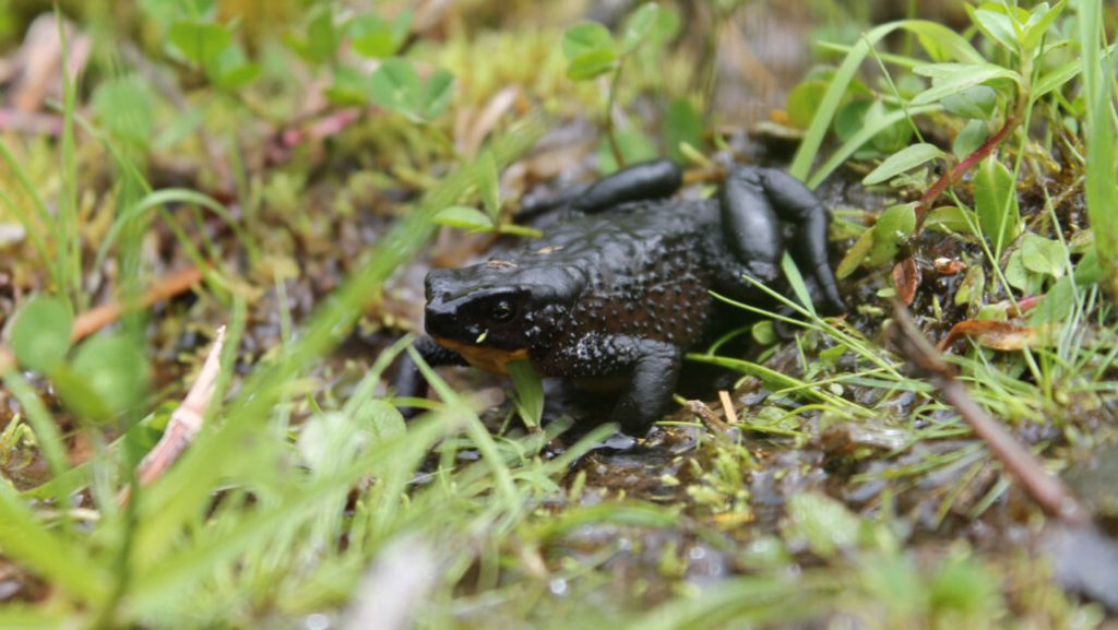
Across Central and South America, one group of bejeweled frogs is making a comeback.
Harlequin frogs — a genus with over 100 brightly colored species — were one of the groups of amphibians hit hardest by a skin-eating chytrid fungus that rapidly spread around the globe in the 1980s (SN: 3/28/19). The group is so susceptible to the disease that with the added pressures of climate change and habitat loss, around 70 percent of known harlequin frog species are now listed as extinct or critically engendered.
But in recent years, roughly one-third of harlequin frogs presumed to have gone extinct since the 1950s have been rediscovered, researchers report in the December Biological Conservation.
The news is a rare “glimmer of hope” in an otherwise bleak time for amphibians around the globe, says Kyle Jaynes, a conservation biologist at Michigan State University in Hickory Corners.
The comeback frog For Jaynes, the path to uncovering how many harlequin frogs have returned from the brink of extinction started when he heard about the Jambato harlequin frog (Atelopus ignescens). This black and orange frog was once so widespread in the Ecuadorian Andes that its common name comes from the word ”jampatu,” which means “frog” in Kichwa, the Indigenous language of the area.
Then came the fungus. From 1988 to 1989, the frogs “just completely disappeared,” Jaynes says. For years, people searched for traces of the frogs. Scientists ran extensive surveys, and pastors offered rewards to their congregants for anyone that could find one.
Then in 2016, a boy discovered a small population of Jambato frogs in a mountain valley in Ecuador. For a species that had been missing for decades, “it seemed like a miracle,” says Luis Coloma, a researcher and conservationist at the Centro Jambatu de Investigación y Conservación de Anfibios in Quito, Ecuador.
Coloma runs a breeding program for Jambato and other Ecuadorian frogs threatened with extinction. In 2019, Jaynes was part of a group of researchers visiting Coloma’s lab to see if they could work out how these frogs had cheated death. After the Jambato frogs returned to the scene, the team started hearing about other missing harlequin species being spotted for the first time in years.
Those stories led Jaynes, Coloma and their colleagues to comb through reports to see just how many harlequin frogs had reappeared. Of the more than 80 species to have gone missing since 1950, as many as 32 species were spotted in the last two decades — a much higher number than the team had expected.
“I think we were all shocked,” Jaynes says.
Ensuring conservation The news comes with caveats. For one thing, it seems like most species avoided disappearing by a hair, and their numbers are still dangerously low. So extinction is still very much on the table. “We’ve got a second chance here,” Jaynes says. “But there is still a lot we have to do to conserve these species.”
Ensuring the continuation of the rediscovered species will depend in part on understanding how they’ve managed to survive so far. Some scientists have speculated that amphibians at higher elevations might be more susceptible to the fungus since it prefers lower temperatures. But a cursory analysis by Jaynes and colleagues revealed that harlequin frogs are being rediscovered at all elevations across their range, indicating that something else may be at play. Jaynes suspects that there is a biological basis for which harlequin frogs live, such as having developed resistance to the fungus (SN: 3/29/18).
Studies like this one can serve as a “launching pad” for understanding how amphibians might survive the dual threats of disease and climate change, says Valerie McKenzie, a disease ecologist at the University of Colorado Boulder who was not involved with the study.
In the meantime, the fact that people are starting to notice the reemergence of species that were once thought to be gone forever “gives me a lot of hope that other species that are harder to observe — because they’re nocturnal or live high in the canopy — are also recovering,” she says. “It motivates me to think we should go look for them.”
Body changes at the brink of adulthood can get awkward in humans, but at least our eyes don’t pop out of our heads on stalks longer than our legs.
High-rise eyes, however, give macho pizzazz to the adult male Pelmatops fruit fly. In one of the stalkier species, P. tangliangi, the eyes-up transformation takes only about 50 minutes, a new study reports. Once stretched, the skinny eyestalks darken and harden, keeping the eyes stuck out like selfie sticks for the rest of the fly’s life. The details of P. tangliangi’s eye lift come from the first published photo sequence of their ocular blossoming, which appears in the September Annals of the Entomological Society of America. Biologists have known that eyestalks evolved in eight different fly families. Yet Pelmatops flies have gotten so little scientific attention that a lot of their basic biology has been a string of question marks.
Video images show the eyestalks curl and rise irregularly. Yet “they are not flopping around while partly inflated,” says Xiaolin Chen, an entomologist and evolutionary biologist at the Chinese Academy of Sciences in Beijing. “They seem slightly stiff, but still flexible enough.”
Females of the species may raise shorter eyestalks too — if Chen and her colleagues have found the right females. Chen suspects that what are now named as two species, based on the few specimens available, may just be two sexes of the same species. The new paper describes a male P. tangliangi mating with a female known by a different species name. Her stalks aren’t as magnificent as his, but she has some.
While the headgear can burden a flying insect, long eyestalks may give flies some swagger. These Pelmatops and other kinds of stalk-eyed flies face off, eyestalk to eyestalk, with uppity intruders. There’s no knocking and locking stalks in fierce fly disputes though. Any pushing and shoving, Chen says, is “done with other body parts.”
Extreme eyes may also have other benefits. In the wild, Chen finds these fruit flies on long stems of Rubus berry brambles. The eyes naturally periscope outward and upward, allowing the flies to spot danger while the body stays hidden in the greenery.
To kill drug-resistant bacteria, “last-resort” antibiotics borrow a tactic from Medusa’s playbook: petrification.
New high-resolution microscope images show that a class of antibiotics called polymyxins crystallize the cell membranes of bacteria. The honeycomb-shaped crystals that form turn the microbes’ usually supple skins of fat molecules into thin brittle sheets, researchers report October 21 in Nature Communications. When the petrified membranes break, the bacteria die.
The finding was a total surprise, says Sebastian Hiller, a structural biologist at the University of Basel in Switzerland. Hiller, biophysicist Selen Manioğlu and their colleagues had been using the antibiotics as a control for a different experiment. When the researchers turned on their microscopes, “we saw these waffles,” Hiller says. “I immediately recognized, wow, this must be something special.”
Polymyxin antibiotics like colistin were discovered in the 1940s and are now used as a powerful last-ditch defense against bacteria that have evolved resistance to most other drugs. Researchers already knew that polymyxins somehow interfere with bacterial cell membranes. But nobody had imagined a scenario like the “waffles” the team discovered. In the new study, Hiller and colleagues exposed bits of cell membrane from Escherichia coli to varying concentrations of colistin. Imaging with atomic force microscopy revealed that crystals formed at the minimum concentrations required to kill the bacteria. Colistin-resistant strains exposed to the drug didn’t form crystals.
The results indicate that polymyxins work by arranging the cell membrane into a crystalline structure that leaves it brittle and vulnerable. “That’s something that has not even remotely been hypothesized so far,” says Markus Weingarth, a biochemist at Utrecht University in the Netherlands who was not involved in the work. “It’s a very important study. I’d even say it’s a breakthrough.”
How exactly polymyxins crystallize cell membranes remains unclear. That’s a problem because some bacteria have developed resistance to polymyxins and are becoming more widespread (SN: 5/27/16; SN: 10/30/90). Without more studies like this one to help reveal how the drugs work, scientists can’t effectively modify the antibiotics to make them more effective, Weingarth says.
Hiller hopes that this first glimpse of polymyxins’ petrifying powers will help scientists combat resistance to the antibiotics.
“Understanding these concepts will definitely bring a lot of ideas — and the potential to make new drugs,” Hiller says.