Bit by qubit, scientists are edging closer to the realm where quantum computers will reign supreme.
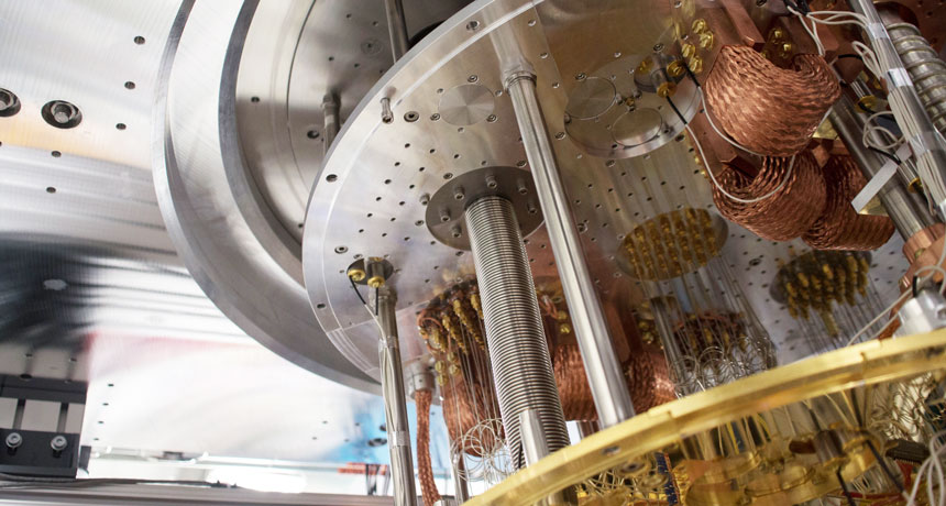
IBM is testing a prototype quantum processor with 50 quantum bits, or qubits, the company announced November 10. That’s about the number needed to meet a sought-after milestone: demonstrating that quantum computers can perform specific tasks that are beyond the reach of traditional computers (SN: 7/8/17, p. 28).
Unlike standard bits, which represent either 0 or 1, qubits can indicate a combination of the two, using what’s called quantum superposition. This property allows quantum computers to perform certain kinds of calculations more quickly. But because qubits are finicky, scaling up is no easy task. Previously, IBM’s largest quantum processor boasted 17 qubits.
IBM also announced a 20-qubit processor that the company plans to make commercially available by the end of the year.
Bit by qubit, scientists are edging closer to the realm where quantum computers will reign supreme.
IBM is now testing a prototype quantum processor with 50 quantum bits, or qubits, the company announced November 10. That’s around the number needed to meet a sought-after milestone: demonstrating that quantum computers can perform specific tasks that are beyond the reach of traditional computers. Unlike standard bits, which represent either 0 or 1, qubits can indicate a combination of the two, using what’s called a quantum superposition. This property allows quantum computers to perform certain kinds of calculations more quickly. But because quantum bits are more finicky than standard bits, scaling up is no easy task. Previously, IBM’s largest quantum processor boasted 17 qubits.
A race is now on to commercialize quantum computers, making them available to companies that want to solve problems particularly suited to quantum machines, such as designing new materials or speeding up the search for new drugs. IBM also announced a 20-qubit processor that the company plans to make commercially available by the end of 2017. Meanwhile, Google has its own plans to commercialize quantum computers. The company’s quantum computing researchers are currently testing a 22-qubit chip and are designing a larger one.
A 26-year-old woman felt something in her left eye. For days, she couldn’t shake the sensation. But this was no errant eyelash or dive-bombing gnat.
A week after that first irritation, the Oregon resident pulled a translucent worm, about a centimeter long, from her eye. With that harrowing feat, she became the first ever reported case of a human infestation with the cattle eyeworm, Thelazia gulosa. “This is a very rare event and exciting from a parasitological perspective,” says medical parasitologist Richard Bradbury of the U.S. Centers for Disease Control and Prevention in Atlanta. “Perhaps not so exciting if you are the patient.” Over 20 days, she and her doctors removed 14 worms from her infected eye, researchers report online February 12 in the American Journal of Tropical Medicine and Hygiene. After that, no more irritation.
T. gulosa is a nematode found in North America, Europe, Australia and central Asia. It infects the large, watchful eyes of cattle. The worm spends its larval stage in the abdomen of the aptly named face fly, Musca autumnalis. As the fly feasts on tears and eye secretions, it spreads the nematode larva, which then grow into adult worms.
Two other Thelazia species are known to infect humans, but rarely. There have been more than 160 cases reported for one species in Europe and Asia, and only 10 cases in North America, by a species found in dogs. This new perpetrator was not expected to be seen in a human, Bradbury says.
The young woman had been horseback riding near cattle farms in Gold Beach, Oregon, which may explain her face-to-face with the fly. “It is just unfortunate for the patient,” Bradbury says, “that she was not able to swish away that one infected fly quickly enough from her eye.”
AUSTIN, Texas — Babies’ stroke-damaged brains can pull a mirror trick to recover.
A stroke on the left side of the brain often damages important language-processing areas. But people who have this stroke just before or after birth recover their language abilities in the mirror image spot on the right side, a study of teens and young adults shows. Those patients all had normal language skills, even though as much as half of their brain had withered away, researchers reported February 17 at the annual meeting of the American Association for the Advancement of Science. Researchers so far have recruited 12 people ages 12 to 25 who had each experienced a stroke to the same region of their brain’s left hemisphere just before or after birth. People who have this type of stroke as adults often lose their ability to use and understand language, said study coauthor Elissa Newport, a neurology researcher at Georgetown University Medical Center in Washington, D.C.
MRI scans of healthy siblings of the stroke patients showed activity in language centers in the left hemisphere of the brain when the participants heard speech. The stroke patients showed activity in the exact same areas — just on the opposite side of the brain.
It’s well established that if an area of the brain gets damaged, other brain areas will sometimes compensate. But the new finding suggests that while young brains have an extraordinary capacity to recover, there might be limits on which areas can pinch-hit.
“When you look at a very well-defined population, recovery takes place in a very particular set of regions,” said Newport. Young children usually show language activity in the same areas on both sides of their brain, Newport noted, and the left side becomes more dominant over time. But in the case of a major stroke to the left side, the corresponding areas on the right side of the brain might already be primed to take over.
Two newly discovered giant viruses have the most comprehensive toolkit for assembling proteins found in any known virus. In a host cell, the viruses have the enzymes needed to wrangle all 20 standard amino acids, the building blocks of life.
Researchers dubbed the viruses Tupanvirus deep ocean and Tupanvirus soda lake, combining the name of the indigenous South American god of thunder, Tupan, with the extreme environment where each type of virus was found. The giant viruses are among the largest of their kind — up to 2.3 micrometers in length — which is about 23 times as long as a particle of HIV, the scientists report February 27 in Nature Communications. Tupanviruses can infect a wide range of hosts, such as protists and amoebas, but pose no threat to humans, the researchers say.
Viruses are considered nonliving, but the genetic complexity of giant viruses has some scientists questioning that categorization. Each Tupanvirus, for example, has a massive genetic instruction book with roughly 1.5 million base pairs of DNA, more than what some bacteria have, says coauthor Bernard La Scola, a virologist at Aix-Marseille University in France.
But other scientists say giant viruses aren’t so different from their smaller kin. Research by Frederik Schulz, with the Department of Energy Joint Genome Institute in Walnut Creek, Calif., suggests these microscopic behemoths are regular viruses that acquired extra genes from hosts and should not be classified as life.
Tupanviruses don’t settle the controversy, but they do challenge our preconceptions of what life is, La Scola says.
Underwater grasses are growing back in the Chesapeake Bay. The plants now carpet three times as much real estate as in 1984, thanks to more than 30 years of efforts to reduce nitrogen pollution. This environmental success story shows that regulations put in place to protect the bay’s health have made a difference, researchers report the week of March 5 in Proceedings of the National Academy of Sciences.
Rules limiting nutrient runoff from farms and wastewater treatment plants helped to decrease nitrogen concentrations in the bay by 23 percent since 1984. That decline in nitrogen has allowed the recovery of 17,000 hectares of grasses, the new study shows — enough to cover roughly 32,000 football fields. “This is one of the best examples we have of linking long-term research data with management to show how important that is in restoring this critical habitat,” says Karen McGlathery, an environmental scientist at the University of Virginia in Charlottesville who wasn’t involved in the research. ”I don’t know of any other system that’s so large and so complicated where these connections have been made.”
The bay’s aquatic vegetation, including seagrasses and freshwater grasses, is an important part of coastal ecosystems, says study coauthor Jonathan Lefcheck, a marine ecologist at the Bigelow Laboratory for Ocean Sciences in East Boothbay, Maine. Beds of underwater grasses act as nurseries that shelter young fish and aquatic invertebrates. The plants clean the water by trapping particulates, and stabilize shorelines by preventing erosion. But the once-lush grasses began dying off in the 1950s when the region’s human population boomed, and cities and farms dumped increasing amounts of nitrogen and other nutrients into the bay.
In the late 1970s and early 1980s, state and federal agencies took action, limiting the amount of nutrients that could enter the bay from farms, water treatment facilities and other sources. Those groups also instituted programs to monitor the bay’s health, building up the stockpile of information that Lefcheck and his colleagues have now analyzed.
The researchers looked at aerial surveys of the bay, data on water temperature and nutrient levels, as well as land and fertilizer use. Using mathematical equations to test which variables had the biggest impact on seagrass regrowth, the team pinned down nitrogen reduction as the driving force. That makes sense: Too much nitrogen in water promotes the growth of plankton, which can block sunlight, and algae, which can settle on the grass blades and smother them. Now, though, researchers are seeing just the opposite. Grasses need clean water to get a foothold, but once they settle in, they “modify their own environment and make it better,” Lefcheck says. “Once you get a little bit established, it can take off.”
As I’ve been reporting a story about the opioid epidemic, I’ve sorted through a lot of tragic numbers that make the astronomical spike in deaths and injuries related to the drugs feel more real.
The rise in the abuse of opioids — powerfully addictive painkillers — is driven by adults. But kids are also swept up in the current, a new study makes clear. The number of children admitted to pediatric intensive care units at hospitals for opioid-related trouble nearly doubled between 2004 and 2015, researchers report in the March Pediatrics. Researchers combed through medical records from 46 hospitals around the United States, looking for opioid-related reasons for admission to the hospital. When the researchers looked at children who landed in pediatric intensive care units for opioid-related crises, the numbers were grim, nearly doubling. In the period including 2004 to 2007, 367 children landed in the PICU for opioid-related trouble. In the period including 2012 to 2015, that number was 643. (From 2008 to 2011, 554 kids were admitted to the PICU for opioid-related illnesses.) Most opioid-related hospital admissions were for children ages 12 to 17, the researchers found. The available stats couldn’t say how many of those events were accidental ingestions versus intentional drug use. (Though for older kids, there’s a sliver of good news from elsewhere: Prescription opioid use among teenagers is actually down, a recent survey suggests.) But about a third of the hospitalizations were for children younger than 6. And among these young kids, about 20 percent of the poisonings involved methadone, a drug that’s used to treat opioid addiction. That means that these young kids are getting into adults’ drugs (illicit or prescribed) and accidentally ingesting them.
Lots of parents don’t store their prescription opioid painkillers safely away from their young children, a survey last year suggests. Drugs, prescription or otherwise, should be kept out of sight and out of reach, ideally locked away. Some kids are great climbers, and some are crafty bottle openers who can, with persistence, work around supposedly child-resistant packaging.
These are tips for everyone living with small kids — not just those who may have opioids in the house. Children are curious, persistent and, sadly, extra vulnerable to powerful drugs, which means that we should all do our best to keep them away from these potentially dangerous drugs.
When you’re pregnant, especially for the first time, you have to make a lot of decisions. Will coffee remain a part of your life? Where are you going to give birth? What are you going to name the baby? What values will you teach him? Do you really need a baby spa bathtub?
Before my first daughter arrived, an instructor at a birth class threw me a curveball: Was I planning on banking my baby’s umbilical cord blood? For much of pregnancy, the umbilical cord is the lifeline of a fetus, tethering it to the placenta. Snaking through the nearly 2-feet-long cord, there’s a vein ferrying nutrients and oxygen from mom’s blood (via the placenta), plus two arteries carrying oxygen- and nutrient-depleted blood from the fetus back to mom. Because mother’s blood and fetal blood don’t actually mix much, the blood in the placenta and umbilical cord at birth belongs mainly to the fetus.
That fetal blood holds all sorts of interesting — and potentially therapeutic — cells and molecules. This realization has, in some cases, changed the way the umbilical cord and placenta are handled during birth. Instead of tossing it aside, some doctors, scientists and parents are choosing to bank this fetal blood — harvesting it from the baby’s umbilical cord and placenta, freezing it and storing it away for later.
Proponents of cord blood banking are convinced that instead of being medical waste, the fetal cells within are biological gold. In this post, and the two that follow, I’ll take a look at the evidence for those claims, and sort through some of the questions that arise as parents consider whether to bank their baby’s cord blood.
Back in the 1980s, umbilical cord blood caught the attention of researchers who suspected that the often-discarded tissue could be a valuable source of shape-shifting stem cells. These cells, which can become several different types of blood cells, are similar to the specialized stem cells found in bone marrow that can churn out new blood cells. Such stem cells are found in adult blood, too, but not as abundantly. In 1988, a 5-year-old named Matthew with a rare type of anemia received umbilical cord blood cells from his newborn sister, who didn’t have the disease. That transfer, called an umbilical cord blood transplant, worked, and the boy was soon free of the disease.
At the time, researchers didn’t know much about the properties of the cells found in umbilical cord blood. But research has zoomed forward, illuminating more about the contents of this young blood.
Of particular interest are the flexible hematopoietic stem cells important in that initial transplant. In certain cases, transplanting these cells might be able to reboot a person’s body and get rid of a disease-related defect. Cord blood transplants are similar to bone marrow transplants. A person with leukemia, for instance, might have his own cancerous blood cells wiped out with chemotherapy and radiation. Healthy, non-cancerous stem cells from a donor can then repopulate the blood.
Extracting stem cells from bone marrow requires surgery under anesthesia; extracting them from the blood requires taking a drug to stimulate their production. And in order to work, these stem cell donations need to come from a person who carries a similar pattern of proteins on the outsides of his or her cells, a molecular calling card known as HLA type. Stem cells found in cord blood don’t need to be as closely matched to work. Because these cells are so flexible, there’s more wiggle room between donor and recipient. That’s particularly good news for people of certain ethnic minorities who often have trouble finding matched stem cell transplant donors.
Hard numbers are tricky to pin down, but between that first transplant in 1988 and 2015, an estimated 35,000 umbilical cord blood transplants had been performed globally. That number includes people treated for leukemia and other types of cancer, blood disorders and immune diseases. And the utility of umbilical cord cells may stretch well beyond the disorders that the cells are currently being used for. “If you read the literature, it’s pretty exciting,” says pediatrician and immunologist William Shearer of Baylor College of Medicine and Texas Children’s Hospital.
Some researchers suspect that umbilical cord blood contains other cells that may have therapeutic effects beyond the blood. Specialized immune cells may be able to tweak brain function, for instance. Trials around the world are studying umbilical cord blood’s capabilities in a wide range of diseases (see Table 2 here): Cerebral palsy, autism, diabetes and lupus are currently under investigation. The cells are even being tested for an ameliorating role in Alzheimer’s disease and other neurodegenerative conditions.
After injections with their own umbilical cord blood, 63 children with cerebral palsy improved on motor skills, on average. And a clinical trial to see whether cord blood transplants improve symptoms of children with autism spectrum disorder should wrap up in the summer of 2018, says pediatric researcher and clinician Joanne Kurtzberg of Duke University, who helped establish a not-for-profit umbilical cord bank in North Carolina. (A small but optimistic pilot study has already been completed.)
The potential powers of these cells have researchers excited. But what that scientific hope means for expectant parents facing decisions about cord blood banking is far from clear. For all of the promise, there are lots of reasons why umbilical cord cells may turn out to be less useful than thought. Read my next post for more about these potential drawbacks.
Greenland is melting rapidly, but some glaciers are disappearing faster than others. A new map of the surrounding seafloor helps explain why: Many of the fastest-melting glaciers sit atop deep fjords that allow Atlantic Ocean water to melt them from below.
Researchers led by glaciologist Romain Millan of the University of California, Irvine analyzed new oceanographic and topographic data for 20 major glaciers within 10 fjords in southeast Greenland. The mapping revealed that some fjords are several hundred meters deeper than simulations of the bathymetry suggested, the researchers report online March 25 in Geophysical Research Letters. These troughs allow warmer and saltier waters from deeper in the ocean to reach the glaciers and erode them. Other glaciers are protected by shallow sills, or raised seafloor ledges. These sills act as barriers to the deep, warm water, the new seafloor maps show. The researchers compared their findings with observations of glacier melt from 1930 to 2017, and found that the fastest-melting glaciers tended to be those more exposed to melting from below. The study uses data from two NASA missions — Operation IceBridge, which measures ice thickness and gravity from aircraft, and Oceans Melting Greenland, or OMG, which uses sonar and gravity instruments to map the shape and depth of the seafloor close to the ice front. The OMG mission also involves dropping hundreds of probes into the ocean each year to measure temperature and salinity at different depths. Scientists have long suspected Greenland’s melting may be accelerated by the ocean (SN Online: 7/6/11), but needed data on fjord depth and glacier thickness to prove it.
The high-resolution OMG datasets, in particular, reveal bumps and troughs in the seafloor that were previously unknown, says glaciologist Andy Aschwanden at the University of Alaska Fairbanks, who was not involved with the study. “Those small details can make quite a difference to when a glacier will retreat.”
Just a few tweaks to a bacterial enzyme make it a lean, mean plastic-destroying machine.
One type of plastic, polyethylene terephthalate, or PET, is widely used in polyester clothing and disposable bottles and is notoriously persistent in landfills. In 2016, Japanese scientists identified a new species of bacteria, Ideonella sakaiensis, which has a specialized enzyme that can naturally break down PET.
Now, an international team of researchers studying the enzyme’s structure has created a variant that’s even more efficient at gobbling plastic, the team reports April 17 in Proceedings of the National Academy of Sciences.
The scientists used a technique called X-ray crystallography to examine the enzyme’s structure for clues to its plastic-killing abilities. Then, they genetically tweaked the enzyme to create small variations in the structure, and tested those versions for PET-degrading performance. Some changes made the enzyme work even better. Both the original version and the mutated versions could break down both PET and another, newer bio-based plastic called PEF, short for polyethylene-2,5-furandicarboxylate. With a little more engineering, these enzymes could someday feast at landfills.