Cows produce powerful HIV antibodies

An unlikely hero has emerged in the quest to fight HIV: the cow. In a first for any animal, including humans, four cows injected with a type of HIV protein rapidly produced powerful antibodies against the virus, researchers report. Learning how to induce similar antibodies in humans may be key to a successful HIV vaccine.

The antibodies, called broadly neutralizing antibodies, can stop infection from a variety of HIV types. The cows generated these antibodies as soon as 42 days after immunization, the researchers report online July 20 in Nature. For the small percentage of people estimated to develop these antibodies after a natural infection, it can take several years.
The work identifies “a new and much more efficient method to generate broadly active antibodies against HIV,” says immunologist Justin Bailey of Johns Hopkins University School of Medicine, who was not involved in the study.

Making an HIV vaccine has proved difficult because the virus changes all the time. Different strains exist throughout the world, and the virus even mutates within an infected person’s body. Most often, people develop antibodies that are specific to one strain but ineffective against others. HIV vaccines tested so far have not led to the production of broadly neutralizing antibodies.

About 1 percent of HIV-infected people eventually generate broadly neutralizing antibodies that are especially potent and effective against many types of HIV. The development of these antibodies doesn’t seem to help infected people. But when given to monkeys before exposure to a virus similar to HIV, the antibodies prevent infection.

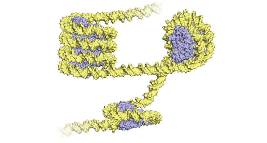
Broadly neutralizing antibodies specific to HIV have a few quirky features, one of which is the presence of a long stretch of amino acids that sticks out from the antibody surface. This protruding part of the antibody binds to a viral site that remains the same between strains, because the virus needs it to gain entry to a cell. HIV’s thick coat of surface sugars makes the viral binding site difficult to access. A longer stretch of amino acids seems to be able to pierce through “and reach in, almost like the long arm of the law,” says Vaughn Smider, a molecular immunologist at the Scripps Research Institute in La Jolla, Calif.

In people infected with HIV who develop broadly neutralizing antibodies, this antibody region — called HCDR3 — has about 30 amino acids, about twice as long as what is usual for human antibodies. Although on the long side for a human, “that’s actually kind of short for a cow,” Smider says.
And so the idea to immunize cows was born. Since cows naturally make longer HCDR3s, Smider explains, perhaps they’d have this sought-after response to HIV.

Smider and colleagues took serum — blood with the cells removed, leaving antibodies behind — from four immunized cows and tested it against different types of HIV virus in a test tube. All of the cows developed broadly neutralizing antibodies. The researchers then tested one cow’s antibodies on an even larger number of virus types. After 381 days, this cow’s antibodies prevented 96 percent of the 117 HIV types from infecting cells in a lab dish. The researchers also isolated an antibody from this cow that had a long HCDR3 of 60 amino acids and stopped infection by 72 percent of the HIV types.

If researchers could induce antibodies with long HCDR3s in humans, “then that could be the basis of getting a vaccine to work,” Smider says. “You need a step before the immunization that helps expand the rare antibodies.” Since cows are so good at making broadly neutralizing antibodies, it also might be possible to turn the cow’s handiwork into drugs for HIV treatment, if bovine antibodies are effective at stopping the virus in other animals, he says.

Half of the Milky Way comes from other galaxies

Galaxies may grow by swiping gas from their neighbors.

New simulations suggest that nearly half the matter in the Milky Way may have been siphoned from the gas of other galaxies. That gas provides the raw material that galaxies use to build their bulk. The finding, scheduled to appear in the Monthly Notices of the Royal Astronomical Society, reveals a new, unexpected way for galaxies to acquire matter and could give clues to how they evolve.
“These simulations show a huge amount of interaction among galaxies, a huge dance that’s going on,” says astronomer Romeel Davé of the University of Edinburgh. That dance, and the subsequent exchange of atoms, could be what establishes a galaxy’s character — whether it’s small or big, elliptical or spiral, quiet or bursting with star formation. If the simulation results are confirmed with observations, it could be a major advancement in understanding galaxy formation, Davé says.

It makes sense that much of the material in one galaxy actually came from other galaxies, says study coauthor Claude-André Faucher-Giguère, a theoretical astrophysicist at Northwestern University in Evanston, Ill. “Still, the result was really unexpected,” he says.

Astronomers thought galaxies got their matter in two main ways. First, atoms clumped together to form stars and then galaxies, not long after the Big Bang about 13.8 billion years ago. Then some of those atoms were eventually ejected by supernovas but rained back onto the same galaxy, recycling the gas again and again.

The new simulations showed a third way galaxies could score gas. Powerful supernova explosions would eject atoms, in the form of gas, far from their home galaxies into intergalactic space. Those atoms would then travel through space, pushed toward other galaxies by galactic winds that move at several hundred kilometers per second. When the particles neared a galaxy’s gravitational pull, they would get sucked in, where they would serve as the basis for stars, planets, dust and other material in their new galactic home. Still, this exchange of atoms is extremely difficult to spot in space because the gas atoms, don’t give off light like stars do.
Faucher-Giguère and colleagues spotted the exchange in computer simulations that show how galaxies formed just after the Big Bang and how they have evolved over time. The team tracked gas atoms as they moved through the model universe, formed stars and then were ejected from galaxies as those stars exploded.

In the simulations, up to half of the atoms in large galaxies were pulled in from other galaxies. Because more massive galaxies have more gravity, they tended to pull atoms from the ejected material of small galaxies. The exchange appears to take billions of years as atoms travel the vast space between galaxies, the team notes.

“It’s that not surprising to see a galaxy kick out matter, which is then pulled in by other galaxies,” Davé says. What is surprising, he says, is the amount of material that’s transferred. Before seeing the simulations, he would have guessed that about 5 percent of gas was transferred among galaxies this way. “To see that it is up to 50 percent is pretty remarkable,” he says.

Already, astronomers are searching for evidence of this material-swapping behavior among galaxies. Faucher-Giguère and colleagues, working with researchers using the Hubble Space Telescope, hope to observe intergalactic transfer of gas among galaxies soon.

The first look at how archaea package their DNA reveals they’re a lot like us

Single-celled microbes may have taught plants and animals how to pack their genetic baggage.

Archaea, a type of single-celled life-form similar to bacteria, keep their DNA wrapped around proteins much in the same way as more complex organisms, researchers report in the Aug. 11 Science. This finding provides new insight into the evolutionary origins of the DNA-packing process and the secret to archaea’s hardiness, which enables some to live in acid, boiling water or other extreme environments.
All eukaryotes, including plants and animals, store their genetic material in cell compartments called nuclei. Such organisms cram meters of genetic material into the tiny nuclei by wrapping strands of DNA around clusters of proteins called histones (SN: 1/10/15, p. 32). “It doesn’t really matter which eukaryote you look at, whether it’s amoebas or plants or humans or fish or insects or anything,” says coauthor John Reeve, a microbiologist at Ohio State University. “They all have exactly the same structure.”

Unlike bacteria, some archaea also contain histones, but researchers weren’t sure whether these microbes spool DNA around the protein bobbins the way eukaryotes do. So Reeve and colleagues used a method called X-ray crystallography to discern, for the first time, the precise shape of archaea DNA bound to histones.

The researchers saw that archaea DNA coils around the histones, similar to the way it does in eukaryotes. “It’s a big deal actually seeing this,” says Steven Henikoff, a molecular biologist at the Fred Hutchinson Cancer Research Center in Seattle who was not involved in the work. The resemblance between archaea and eukaryote DNA wrapping means that the first organism that used this storage scheme was an ancestor of both modern eukaryotes and archaea, the researchers conclude.

But the way archaea DNA twists around histones isn’t identical to the coils of DNA seen in eukaryotes. In eukaryotes, a strand of DNA loops twice around a cluster of eight histones to create what’s called a nucleosome, and connects many of these nucleosomes like beads on a string. Archaea DNA string together bundles of proteins, too. But while eukaryotes always tether eight-protein clumps, archaea DNA can spiral around stacks of many more histones to create rod-shaped structures of various lengths. “So it’s not as uniform as in eukaryotes,” says coauthor Karolin Luger, a biophysicist and Howard Hughes Medical Institute investigator at the University of Colorado Boulder.

Researchers tested the importance of that rodlike architecture by tampering with the histone-DNA structures of some archaea and then observing how these mutant archaea fared in different conditions. “We tried to mimic some real-life situations that some of these organisms could get into,” Luger says.
For instance, some archaea that live in volcanic vents that emit sulfurous gases sometimes get spewed out and have to survive sans sulfur. Archaea with normal histone-DNA shapes can handle that kind of midlife crisis. But when researchers cut their mutant microbes off sulfur, the microorganisms’ growth was stunted. These microbes may not have been able to adapt to sulfur deprivation as well as their wild counterparts “because they can’t unpackage their DNA as readily if the structure has been changed,” Reeve says.

Henikoff calls it “a pretty cool experiment.” It showed that the archaea’s particular DNA-histone architecture was “biologically relevant, not just a novelty,” he says.

Eclipse watchers will go after the biggest solar mystery: Why is the corona so hot?

A total solar eclipse shines a light on the sun’s elusive atmosphere. When the moon blocks the sun, it’s finally possible to see how this diffuse cloud of plasma, called the corona, is magnetically sculpted into beautiful loops. The material there is about a trillionth the density of the solar surface. From its delicate and diaphanous appearance, you might expect the corona to be where the sun goes to cool off.

That couldn’t be more wrong. The corona is a mysteriously sizzling inferno where the temperature jumps from a mere few thousand degrees to several million degrees. Why?
“It’s one of the longest unanswered questions in all of solar physics,” says Paul Bryans of the High Altitude Observatory at the National Center for Atmospheric Research in Boulder, Colo. “There are a bunch of different ideas about what’s going on there, but it’s still highly debated.” Data collected during the Aug. 21 solar eclipse may bring scientists closer to settling that debate.

The sun simmers at about 5,500° Celsius at its visible surface, the photosphere. But the gas just above the photosphere is heated to about 10,000° C. Then in the corona, the temperature makes an abrupt jump to several million degrees.

“It’s counterintuitive that as you move away from a heat source, it gets warmer,” Bryans says. The corona’s diffuseness makes its heat even stranger — the most basic ways to heat a material rely on particles crashing into each other, but the corona is too tenuous for that to work.

An eclipse first brought this abnormal arrangement to light. German astronomer Walter Grotrian observed spectral lines — the fingerprints of elements that show up when light is split into its component wavelengths — emitted by the corona during a total solar eclipse in 1869.

Astronomers at first assumed those lines were due to a new element they dubbed coronium. But Grotrian realized that iron atoms stripped of several of their electrons by the heat were responsible. These iron lines in the corona are still used to measure its temperature: The more electrons lost, the hotter the material in the corona (SN Online: 6/16/17).
Such extreme temperatures have something to do with the corona’s magnetic field, which is probably where all that energy is stored. Once the energy is there, the corona has a hard time radiating it away, so it builds up. Most of the ways that materials release energy — stripping electrons from atoms, accelerating those electrons so they release X-rays and ultraviolet particles of light — are already maxed out in the corona.
“We know there’s energy coming in, and it’s hard to get it out unless you get very hot,” says Amir Caspi of the Southwest Research Institute in Boulder, Colo. “What we don’t understand is how that energy gets into the corona in the first place.”

Physicists have several ideas. Maybe loops of magnetic field lines in the corona vibrate like guitar strings, heating things up, sort of like how a microwave oven heats food. Maybe the magnetic anchors of those loops on the sun’s surface braid and twist the magnetic field above them, dumping in energy that is then continually radiated away like the heating element in a toaster.

Or maybe tiny explosions called nanoflares or jets called spicules carry energy away from the photosphere and into the corona. The formation of new coronal loops that connect to existing ones could dump in enough extra energy to heat the plasma up.

During the solar eclipse, dozens of groups of scientists across the country will deploy telescopes equipped with filters to pick out polarized light, infrared light or those electron-deprived iron atoms in search of answers. Bryans and his colleagues will be on a mountaintop near Casper, Wyo., in the path of totality. There, the team will take images at a fast clip in both visual and infrared wavelengths to map how the corona changes as the moon moves across the sun. (I will be in Wyoming with this team on the day of the eclipse and will be sharing more about how the experiments went.)

“We can look at how things change as we move from the surface up into the atmosphere,” Bryans says. “How that changes is tied to understanding how the corona is heated.”

Probably all of those mechanisms scientists have thought up contribute to the corona’s extreme heat. It’s difficult to declare just one the most important. But ultimately, the solar eclipse is the best chance scientists have to test them. It’s the only time the corona is the star of the solar show.

How horses lost their toes

Horses can leap over high hurdles, gallop at speeds of up to 70 kilometers per hour and haul around up to nearly 1,000 kilograms of body weight — and all with just one big toe on each foot. Now, a new study published August 23 in Proceedings of the Royal Society B helps explain why: Streamlined digits improved horses’ strength and speed.

Along with zebras and donkeys, horses are among the few single-toed creatures in the animal kingdom. Scientists have long suspected that horses’ single, hoofed toes helped them run farther and faster over grasslands, letting them flee predators and find fresh forage. But the hypothesis that having one big toe is better than having several, biomechanically speaking, has never been directly tested.
“This study takes an important step” toward resolving why horses shed digits during their early evolution, says Karen Sears, an evolutionary biologist at UCLA.

Ancient horses had a lot of toes to lose. The dog-sized Hyracotherium, which lived about 55 million years ago, had four toes on its front feet, and three on its back feet. Merychippus, which lived about 10 million years ago, resembled a modern horse but had three toes, including one long middle digit with a protective, toenail-like hoof at the end. The only surviving horse genus, single-toed Equus, emerged about 5 million years ago.

“If you look closely, you can still see the vestigial remnants” of a bone that would have led to a side toe on a modern horse’s foot, says Brianna McHorse, a paleontologist at Harvard University.

To retrace the evolution of horse toes, McHorse and colleagues used CT scans to capture the internal structure of fossilized foot bones from 12 kinds of extinct horses. They also analyzed the feet of the closely related Central American tapir, which are oddly toed like Hyracotherium. A computer simulation then let researchers estimate how the bones would respond to the stresses of locomotion for each species, such as jumping over a hurdle or accelerating into a gallop. Then the scientists compared what happened when they applied the animal’s full body weight to just to the central toe, or spread it among multiple toes.

Side toes significantly increased the early horses’ ability to bear their own weight, the team found — the central toe of early horses would have fractured without help from other toes. As the era of modern horses approached and side toes dropped away, however, the middle toe bone grew thicker and hollower. These changes made the single-toed foot nearly as sturdy — resistant to bending and compression — as multiple toes.
As horses’ legs grew longer, the extra toes at the end of the limb would have been “like wearing weights around your ankles,” McHorse says. Shedding those toes could have helped early horses save energy, allowing them to travel farther and faster, she says. The study can’t determine what changes came first — whether bulking up the middle toe drove the loss of side toes, or the loss of side toes caused changes in the middle toe.

Horses aren’t the only animals to have lost toes or fingers to the evolutionary chopping block. “Digits have been lost many times in animals that walk, run, hop and fly,” says Kim Cooper, a biologist at the University of California, San Diego. Modeling how forces of locomotion act on an animal’s bones — living or extinct — could help scientists understand why.

Brain chemical lost in Parkinson’s may contribute to its own demise

The brain chemical missing in Parkinson’s disease may have a hand in its own death. Dopamine, the neurotransmitter that helps keep body movements fluid, can kick off a toxic chain reaction that ultimately kills the nerve cells that make it, a new study suggests.

By studying lab dishes of human nerve cells, or neurons, derived from Parkinson’s patients, researchers found that a harmful form of dopamine can inflict damage on cells in multiple ways. The result, published online September 7 in Science, “brings multiple pieces of the puzzle together,” says neuroscientist Teresa Hastings of the University of Pittsburgh School of Medicine.
The finding also hints at a potential treatment for the estimated 10 million people worldwide with Parkinson’s: Less cellular damage occurred when some of the neurons were treated early on with antioxidants, molecules that can scoop up harmful chemicals inside cells.

Study coauthor Dimitri Krainc, a neurologist and neuroscientist at Northwestern University Feinberg School of Medicine in Chicago, and colleagues took skin biopsies from healthy people and people with one of two types of Parkinson’s disease, inherited or spontaneously arising. The researchers then coaxed these skin cells into becoming dopamine-producing neurons. These cells were similar to those found in the substantia nigra, the movement-related region of the brain that degenerates in Parkinson’s.
After neurons carrying a mutation that causes the inherited form of Parkinson’s had grown in a dish for 70 days, the researchers noticed some worrisome changes in the cells’ mitochondria. Levels of a harmful form of dopamine known as oxidized dopamine began rising in these energy-producing organelles, reaching high levels by day 150. Neurons derived from people with the more common, sporadic form of Parkinson’s showed a similar increase but later, beginning at day 150. Cells derived from healthy people didn’t accumulate oxidized dopamine.
This dangerous form of dopamine seemed to kick off other types of cellular trouble. Defects in the cells’ lysosomes, cellular cleanup machines, soon followed. So did the accumulation of a protein called alpha-synuclein, which is known to play a big role in Parkinson’s disease.
Those findings are “direct experimental evidence from human cells that the very chemical lost in Parkinson’s disease contributes to its own demise,” says analytical neurochemist Dominic Hare, of the Florey Institute of Neuroscience and Mental Health in Melbourne, Australia. Because these cells churn out dopamine, they are more susceptible to dopamine’s potential destructive forces, he says.

When researchers treated neurons carrying a mutation that causes inherited Parkinson’s with several different types of antioxidants, the damage was lessened. To work in people, antioxidants would need to cross the blood-brain barrier, a difficult task, and reach the mitochondria in the brain. And this would need to happen early, probably even before symptoms appear, Krainc says.

“Without this human model, we would not have been able to untangle the pathway,” Krainc says. In dishes of mouse neurons with Parkinson’s-related mutations, dopamine didn’t kick off the same toxic cascade, a difference that might be due to human neurons containing more dopamine than mice neurons. Dopamine-producing neurons in mice and people “have some very fundamental differences,” Krainc says. And those differences might help explain why discoveries in mice haven’t translated to treatments for people with Parkinson’s, he says.

Over the past few decades, scientists have been accumulating evidence that oxidized dopamine can contribute to Parkinson’s disease, Hastings says. Given that knowledge, the new results are expected, she says, but still welcome confirmation of the idea.

These toxic cellular events occurred in lab dishes, not actual brains. “Cell cultures aren’t the perfect re-creation of what’s going on in the human brain,” Hare cautions. But these types of experiments are “the next best thing for monitoring the chemical changes” in these neurons, he says.

This giant marsupial was a seasonal migrant

The largest marsupial to ever walk the Earth just got another accolade: It’s also the only marsupial known to migrate seasonally.

Diprotodon optatum was a massive wombat-like herbivore that lived in what’s now Australia and New Guinea during the Pleistocene, until about 40,000 years ago. Now, an analysis of one animal’s teeth suggests that it undertook long, seasonal migrations like those made by zebras and wildebeests in Africa.

Animals pick up the chemical element strontium through their diet, and it leaves a record in their teeth. The ratio of different strontium isotopes varies from place to place, so it can provide clues about where an animal lived. Strontium isotope ratios in an incisor from one D. optatum revealed a repeating pattern. That suggests the animal migrated seasonally — it moved around, but generally hit up the same rest stops each year, researchers report September 27 in the Proceedings of the Royal Society B.

It’s the first evidence to show a marsupial — living or extinct — migrating in this way, says study coauthor Gilbert Price, a paleoecologist at the University of Queensland in Brisbane, Australia. It’s not clear exactly why this mega-marsupial might have migrated, but an analysis of the carbon isotopes in its teeth suggests it ate a fairly limited diet. So it might have migrated to follow food sources that popped up seasonally in different places, the authors suggest.

14 cattle eyeworms removed from Oregon woman’s eye

A 26-year-old woman felt something in her left eye. For days, she couldn’t shake the sensation. But this was no errant eyelash or dive-bombing gnat.

A week after that first irritation, the Oregon resident pulled a translucent worm, about a centimeter long, from her eye. With that harrowing feat, she became the first ever reported case of a human infestation with the cattle eyeworm, Thelazia gulosa. “This is a very rare event and exciting from a parasitological perspective,” says medical parasitologist Richard Bradbury of the U.S. Centers for Disease Control and Prevention in Atlanta. “Perhaps not so exciting if you are the patient.”
Over 20 days, she and her doctors removed 14 worms from her infected eye, researchers report online February 12 in the American Journal of Tropical Medicine and Hygiene. After that, no more irritation.

T. gulosa is a nematode found in North America, Europe, Australia and central Asia. It infects the large, watchful eyes of cattle. The worm spends its larval stage in the abdomen of the aptly named face fly, Musca autumnalis. As the fly feasts on tears and eye secretions, it spreads the nematode larva, which then grow into adult worms.

Two other Thelazia species are known to infect humans, but rarely. There have been more than 160 cases reported for one species in Europe and Asia, and only 10 cases in North America, by a species found in dogs. This new perpetrator was not expected to be seen in a human, Bradbury says.

The young woman had been horseback riding near cattle farms in Gold Beach, Oregon, which may explain her face-to-face with the fly.
“It is just unfortunate for the patient,” Bradbury says, “that she was not able to swish away that one infected fly quickly enough from her eye.”

Babies can recover language skills after a left-side stroke

AUSTIN, Texas — Babies’ stroke-damaged brains can pull a mirror trick to recover.

A stroke on the left side of the brain often damages important language-processing areas. But people who have this stroke just before or after birth recover their language abilities in the mirror image spot on the right side, a study of teens and young adults shows. Those patients all had normal language skills, even though as much as half of their brain had withered away, researchers reported February 17 at the annual meeting of the American Association for the Advancement of Science.
Researchers so far have recruited 12 people ages 12 to 25 who had each experienced a stroke to the same region of their brain’s left hemisphere just before or after birth. People who have this type of stroke as adults often lose their ability to use and understand language, said study coauthor Elissa Newport, a neurology researcher at Georgetown University Medical Center in Washington, D.C.

MRI scans of healthy siblings of the stroke patients showed activity in language centers in the left hemisphere of the brain when the participants heard speech. The stroke patients showed activity in the exact same areas — just on the opposite side of the brain.

It’s well established that if an area of the brain gets damaged, other brain areas will sometimes compensate. But the new finding suggests that while young brains have an extraordinary capacity to recover, there might be limits on which areas can pinch-hit.

“When you look at a very well-defined population, recovery takes place in a very particular set of regions,” said Newport. Young children usually show language activity in the same areas on both sides of their brain, Newport noted, and the left side becomes more dominant over time. But in the case of a major stroke to the left side, the corresponding areas on the right side of the brain might already be primed to take over.

These giant viruses have more protein-making gear than any known virus

Two newly discovered giant viruses have the most comprehensive toolkit for assembling proteins found in any known virus. In a host cell, the viruses have the enzymes needed to wrangle all 20 standard amino acids, the building blocks of life.

Researchers dubbed the viruses Tupanvirus deep ocean and Tupanvirus soda lake, combining the name of the indigenous South American god of thunder, Tupan, with the extreme environment where each type of virus was found. The giant viruses are among the largest of their kind — up to 2.3 micrometers in length — which is about 23 times as long as a particle of HIV, the scientists report February 27 in Nature Communications.
Tupanviruses can infect a wide range of hosts, such as protists and amoebas, but pose no threat to humans, the researchers say.

Viruses are considered nonliving, but the genetic complexity of giant viruses has some scientists questioning that categorization. Each Tupanvirus, for example, has a massive genetic instruction book with roughly 1.5 million base pairs of DNA, more than what some bacteria have, says coauthor Bernard La Scola, a virologist at Aix-Marseille University in France.

But other scientists say giant viruses aren’t so different from their smaller kin. Research by Frederik Schulz, with the Department of Energy Joint Genome Institute in Walnut Creek, Calif., suggests these microscopic behemoths are regular viruses that acquired extra genes from hosts and should not be classified as life.

Tupanviruses don’t settle the controversy, but they do challenge our preconceptions of what life is, La Scola says.