Narrowed plumbing lets flower survive summer cold snaps
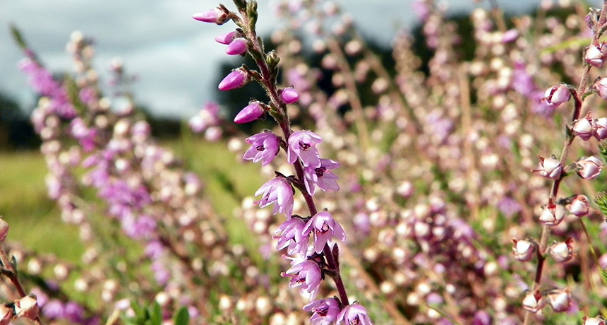
A summertime cold snap can, quite literally, take the bloom off the rose. Not so for Scotch heather — and now scientists know why.
Thick cell walls and narrow plumbing in the alpine shrub’s stems stop deadly ice crystals from spreading to its fragile flowers during sudden summer freezes, researchers report September 15 in PLOS ONE. That lets the flowers survive and the plant make seeds even if temperatures dip below freezing.
Once ice crystals start to form inside of a plant, they can spread very quickly, says Gilbert Neuner, a botanist at the University of Innsbruck in Austria who led the study. Those sharp crystals can destroy plant cells — and flowers are particularly sensitive. So plants living in cold climes have developed strategies to confine ice damage to less harmful spots.
Neuner and his team used infrared imaging to measure heat given off by Scotch heather (Calluna vulgaris) plants as they freeze. That technique revealed where and when ice was forming. And looking at thin slices of the plant under a microscope let the scientists pick apart the structure of the plant’s ice barrier.
Cells at the base of the flower stalks had thicker walls and were packed more closely together than elsewhere in the plant, the team found. In the same area, the pipelines that carry water up the plant — called xylem — were narrower and had fewer points where ice could potentially sneak through. Those modifications let the plants “supercool” their flowers. That is, even when the flowers chilled to below zero degrees Celsius, they contained liquid water instead of ice. Ice didn’t form in the Scotch heather flowers until far below normal freezing temperatures, ‒22° C, and ice that formed elsewhere in the plant didn’t spread to the flowers.
Other species sometimes put up temporary ice blockades, for instance to protect overwintering buds. But that usually cuts off the flow of water through the xylem — fine if a plant is dormant over the winter, but flowers facing a sudden summer freeze need a continuous supply of water. Scotch heather gets around this problem by threading its xylem right through the icy barrier.
Membranes let water pass between the xylem cells, and these membranes might ultimately control the spread of ice crystals in C. vulgaris, Neuner suspects. Tiny pores in the membranes are too small to let ice crystals through the barrier. And when water molecules are found inside such small holes, the molecules are bound so tightly to the structures around them that they behave more like a gel instead of crystalizing into ice. The team hopes to test the idea in future studies.
Other flowering alpine plants could use a similar strategy. “I don’t think that this is unique to this plant,” says Sanna Sevanto, a tree physiologist at Los Alamos National Laboratory in New Mexico who wasn’t involved in the study. “It’s just that nobody has looked at it.”