Brain chemical lost in Parkinson’s may contribute to its own demise
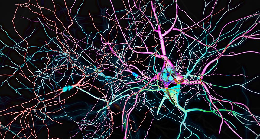
The brain chemical missing in Parkinson’s disease may have a hand in its own death. Dopamine, the neurotransmitter that helps keep body movements fluid, can kick off a toxic chain reaction that ultimately kills the nerve cells that make it, a new study suggests.

By studying lab dishes of human nerve cells, or neurons, derived from Parkinson’s patients, researchers found that a harmful form of dopamine can inflict damage on cells in multiple ways. The result, published online September 7 in Science, “brings multiple pieces of the puzzle together,” says neuroscientist Teresa Hastings of the University of Pittsburgh School of Medicine.
The finding also hints at a potential treatment for the estimated 10 million people worldwide with Parkinson’s: Less cellular damage occurred when some of the neurons were treated early on with antioxidants, molecules that can scoop up harmful chemicals inside cells.

Study coauthor Dimitri Krainc, a neurologist and neuroscientist at Northwestern University Feinberg School of Medicine in Chicago, and colleagues took skin biopsies from healthy people and people with one of two types of Parkinson’s disease, inherited or spontaneously arising. The researchers then coaxed these skin cells into becoming dopamine-producing neurons. These cells were similar to those found in the substantia nigra, the movement-related region of the brain that degenerates in Parkinson’s.
After neurons carrying a mutation that causes the inherited form of Parkinson’s had grown in a dish for 70 days, the researchers noticed some worrisome changes in the cells’ mitochondria. Levels of a harmful form of dopamine known as oxidized dopamine began rising in these energy-producing organelles, reaching high levels by day 150. Neurons derived from people with the more common, sporadic form of Parkinson’s showed a similar increase but later, beginning at day 150. Cells derived from healthy people didn’t accumulate oxidized dopamine.
This dangerous form of dopamine seemed to kick off other types of cellular trouble. Defects in the cells’ lysosomes, cellular cleanup machines, soon followed. So did the accumulation of a protein called alpha-synuclein, which is known to play a big role in Parkinson’s disease.
Those findings are “direct experimental evidence from human cells that the very chemical lost in Parkinson’s disease contributes to its own demise,” says analytical neurochemist Dominic Hare, of the Florey Institute of Neuroscience and Mental Health in Melbourne, Australia. Because these cells churn out dopamine, they are more susceptible to dopamine’s potential destructive forces, he says.

When researchers treated neurons carrying a mutation that causes inherited Parkinson’s with several different types of antioxidants, the damage was lessened. To work in people, antioxidants would need to cross the blood-brain barrier, a difficult task, and reach the mitochondria in the brain. And this would need to happen early, probably even before symptoms appear, Krainc says.

“Without this human model, we would not have been able to untangle the pathway,” Krainc says. In dishes of mouse neurons with Parkinson’s-related mutations, dopamine didn’t kick off the same toxic cascade, a difference that might be due to human neurons containing more dopamine than mice neurons. Dopamine-producing neurons in mice and people “have some very fundamental differences,” Krainc says. And those differences might help explain why discoveries in mice haven’t translated to treatments for people with Parkinson’s, he says.

Over the past few decades, scientists have been accumulating evidence that oxidized dopamine can contribute to Parkinson’s disease, Hastings says. Given that knowledge, the new results are expected, she says, but still welcome confirmation of the idea.

These toxic cellular events occurred in lab dishes, not actual brains. “Cell cultures aren’t the perfect re-creation of what’s going on in the human brain,” Hare cautions. But these types of experiments are “the next best thing for monitoring the chemical changes” in these neurons, he says.

3-D scans of fossils suggest new fish family tree

When it comes to some oddball fish, looks can be deceiving.

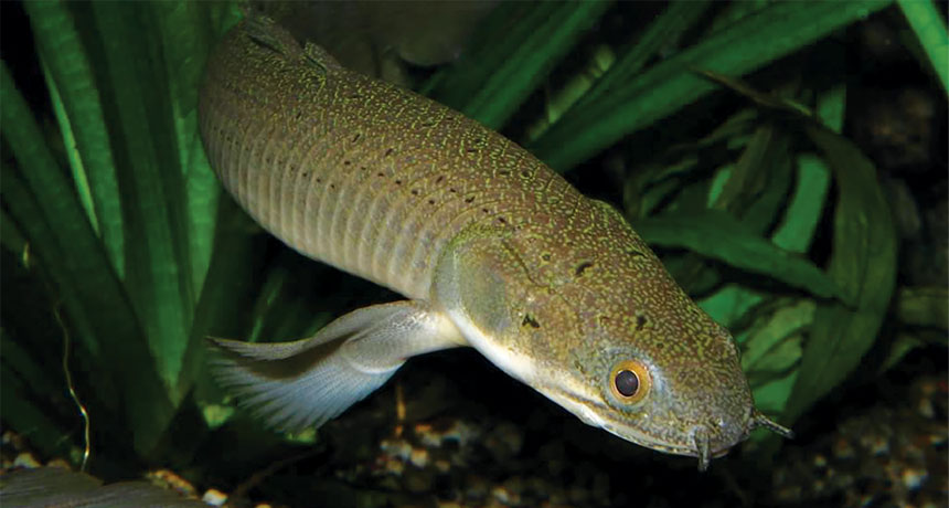
Polypterus, today found only in Africa, and its close kin have generally been considered some of the most primitive ray-finned fishes alive, thanks in part to skeletal features that resemble those on some ancient fish. Now a new analysis of fish fossils of an early polypterid relative called Fukangichthys unearthed in China suggests that those features aren’t so old. The finding shakes up the evolutionary tree of ray-finned fishes, making the group as a whole about 20 million to 40 million years younger than thought, researchers propose online August 30 in Nature.
Ray-finned fishes named for the spines, or rays, that support their fins — are the largest group of vertebrates, making up about half of all backboned animals. They include 30,000 living species, such as gars, bowfins and salmon. The group was thought to originate about 385 million years ago, in the Devonian Period. But the new research, using 3-D CT scans of the previously discovered fossils, shifts the fishes’ apparent origin to the start of the Carboniferous Period some 360 million years ago, says study coauthor Matt Friedman, a paleontologist at the University of Michigan in Ann Arbor.
One of the largest extinction events in Earth’s history marks the boundary between the Devonian and Carboniferous. “We know that many groups of backboned animals were hard hit by the event,” Friedman says. But after the massive die-off, ray-finned fishes popped up and, according to previous fossil evidence, their diversity exploded. The new finding “brings the origin of the modern ray-finned fish group in line with this conspicuous pattern that we see in the fossil record,” Friedman says. It suggests these vertebrates didn’t survive the event. They came after, then flourished.

Why it’s good news that Pluto doesn’t have rings

Pluto has no rings — New Horizons triple-checked. An exhaustive search for rings and dust particles around the dwarf planet before, during and after the spacecraft flew past Pluto in 2015 has come up empty.

“It’s a very long paper to say we didn’t find anything,” says team member Tod Lauer of the analysis, posted online September 23 at arXiv.org. But the nonresult could help scientists understand the contents of the outer solar system — and help plan New Horizons’ next encounter. The spacecraft is now on a course to a space rock in the Kuiper Belt, another 1.5 billion kilometers past Pluto.
Before New Horizons arrived at Pluto, the possible existence of rings was an urgent matter of safety. Hitting a particle as small as a sand grain could have damaged the spacecraft.

Searches with the Hubble Space Telescope in 2011 and 2012 turned up two previously unknown moons orbiting Pluto — Kerberos and Styx (SN: 11/28/15, p. 14) — and zero rings. Even so, many researchers expected to encounter rings, or at least some debris. The four outer planets in the solar system have rings, as do other small bodies in the solar system, like the tiny planetoid 10199 Chariklo (SN: 5/3/14, p. 10). And some studies suggest that Pluto probably had rings at one point in its past, left over from the collision that formed its largest moon, Charon.

Nine weeks before New Horizons’ closest approach to Pluto, a team jokingly called the “crow’s nest” acted much like a ship’s lookout for potential hazards, says Lauer, an astronomer with the National Optical Astronomy Observatory in Tucson, Ariz. The group examined images taken with the spacecraft’s Long Range Reconnaissance Imager camera, looking for ring particles reflecting sunlight or spots that moved against a starry background from one set of images to the next. Nothing turned up.

The team declared the spacecraft’s trajectory safe, and New Horizons flew sailed safely past Pluto on July 14, 2015 (SN Online: 7/15/15). After the flyby, the team turned New Horizons around to look back at Pluto, and towards the sun. This was a much better position to look for rings, as dust particles would pop into view when backlit by the sun like motes of dust in the light from a window.

“If you really want to know for sure whether there’s any dust there, the viewing geometries where you’re looking past the dust with the sun in the background, that’s the gold standard,” says Matthew Tiscareno of the SETI Institute in Mountain View, Calif., who studied Saturn’s rings with the Cassini spacecraft but was not involved in New Horizons.
It took the better part of a year for all the data from New Horizons to return to Earth, and several months after that to analyze it, but the team is now ready to call it: The rings really aren’t there — or at least they’re too diffuse to see.

That’s somewhat surprising, Lauer says. But the chaotic gravity of Pluto’s family of moons might make it too hard for rings to find stable orbits. Or the slight pressure generated by light particles streaming from the sun could constantly blow would-be ring particles away.

It’s also possible there just wasn’t that much dust there to begin with. New Horizons saw fewer craters on Pluto and Charon than expected, which could mean there are fewer small bodies at that distance from the sun smacking into Pluto and its moons and kicking up dust.

That could be good news for New Horizons’ next act. After five months in hibernation, the spacecraft woke up on September 11 and has set its sights on a smaller, weirder and more distant object: a space rock about 30 kilometers long called 2014 MU69 (SN Online: 7/20/17). Initial observations suggest it might be a double object, with two bodies orbiting closely or touching lightly.

New Horizons will fly past MU69 on January 1, 2019. In the meantime, the team is looking for hazards along the route. “We’re going to do a similar effort to what we did with Pluto,” Lauer says. “We’re going to get in the crow’s nest and get out our binoculars, as it were, and see if we’re going to be okay.”

An interstellar asteroid might have just been spotted for the first time

Astronomers may have just spotted the first asteroid caught visiting the solar system from another star.

The Pan-STARRS 1 telescope in Hawaii discovered the object, initially dubbed A/2017 U1 and later named ‘Oumuamua, on October 18. More observations from other telescopes around the world suggest the object’s trajectory is at an unusually steep angle to the plane on which all the planets lie, and it does not orbit the sun. A/2017 U1’s slingshot route suggests it is a recent visitor to the solar system — and is now on its way out again. The discovery was announced in a bulletin published October 25 by the International Astronomical Union’s Minor Planet Center.
All asteroids previously seen come from within the solar system and circle the sun. Even comets, which come from a distant reservoir of icy rocks in the solar system called the Oort cloud and can have highly titled orbits, still orbit the sun.

Astronomers first pegged the object as a comet thanks to its elongated path, but additional telescope observations October 25 indicate it’s more likely that A/2017 U1 is an asteroid. Those observations revealed that the object looked like a single, sharp point of light, suggesting it is not a comet, which would have an extended icy halo. The asteroid, which is probably no more than 400 meters across, zoomed into the solar system at 25.5 kilometers per second and is now fleeing at 44 km/s.

The new data also supported the wacky trajectory, suggesting the object truly is a visitor from beyond. “It’s now looking very promising,” says planetary scientist Michele Bannister of Queen’s University Belfast in Northern Ireland, although she would still like to get more data to be sure. Astronomers are already planning to measure the colors in the asteroid’s reflected light to figure out what it’s made of, a clue to its origins.

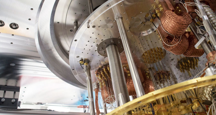
Quantum computing steps forward with 50-qubit prototype

Bit by qubit, scientists are edging closer to the realm where quantum computers will reign supreme.

IBM is testing a prototype quantum processor with 50 quantum bits, or qubits, the company announced November 10. That’s about the number needed to meet a sought-after milestone: demonstrating that quantum computers can perform specific tasks that are beyond the reach of traditional computers (SN: 7/8/17, p. 28).

Unlike standard bits, which represent either 0 or 1, qubits can indicate a combination of the two, using what’s called quantum superposition. This property allows quantum computers to perform certain kinds of calculations more quickly. But because qubits are finicky, scaling up is no easy task. Previously, IBM’s largest quantum processor boasted 17 qubits.

IBM also announced a 20-qubit processor that the company plans to make commercially available by the end of the year.

Quantum computers take a step forward with a 50-qubit prototype

Bit by qubit, scientists are edging closer to the realm where quantum computers will reign supreme.

IBM is now testing a prototype quantum processor with 50 quantum bits, or qubits, the company announced November 10. That’s around the number needed to meet a sought-after milestone: demonstrating that quantum computers can perform specific tasks that are beyond the reach of traditional computers. Unlike standard bits, which represent either 0 or 1, qubits can indicate a combination of the two, using what’s called a quantum superposition. This property allows quantum computers to perform certain kinds of calculations more quickly. But because quantum bits are more finicky than standard bits, scaling up is no easy task. Previously, IBM’s largest quantum processor boasted 17 qubits.

A race is now on to commercialize quantum computers, making them available to companies that want to solve problems particularly suited to quantum machines, such as designing new materials or speeding up the search for new drugs. IBM also announced a 20-qubit processor that the company plans to make commercially available by the end of 2017. Meanwhile, Google has its own plans to commercialize quantum computers. The company’s quantum computing researchers are currently testing a 22-qubit chip and are designing a larger one.

14 cattle eyeworms removed from Oregon woman’s eye

A 26-year-old woman felt something in her left eye. For days, she couldn’t shake the sensation. But this was no errant eyelash or dive-bombing gnat.

A week after that first irritation, the Oregon resident pulled a translucent worm, about a centimeter long, from her eye. With that harrowing feat, she became the first ever reported case of a human infestation with the cattle eyeworm, Thelazia gulosa. “This is a very rare event and exciting from a parasitological perspective,” says medical parasitologist Richard Bradbury of the U.S. Centers for Disease Control and Prevention in Atlanta. “Perhaps not so exciting if you are the patient.”
Over 20 days, she and her doctors removed 14 worms from her infected eye, researchers report online February 12 in the American Journal of Tropical Medicine and Hygiene. After that, no more irritation.

T. gulosa is a nematode found in North America, Europe, Australia and central Asia. It infects the large, watchful eyes of cattle. The worm spends its larval stage in the abdomen of the aptly named face fly, Musca autumnalis. As the fly feasts on tears and eye secretions, it spreads the nematode larva, which then grow into adult worms.

Two other Thelazia species are known to infect humans, but rarely. There have been more than 160 cases reported for one species in Europe and Asia, and only 10 cases in North America, by a species found in dogs. This new perpetrator was not expected to be seen in a human, Bradbury says.

The young woman had been horseback riding near cattle farms in Gold Beach, Oregon, which may explain her face-to-face with the fly.
“It is just unfortunate for the patient,” Bradbury says, “that she was not able to swish away that one infected fly quickly enough from her eye.”

Babies can recover language skills after a left-side stroke

AUSTIN, Texas — Babies’ stroke-damaged brains can pull a mirror trick to recover.

A stroke on the left side of the brain often damages important language-processing areas. But people who have this stroke just before or after birth recover their language abilities in the mirror image spot on the right side, a study of teens and young adults shows. Those patients all had normal language skills, even though as much as half of their brain had withered away, researchers reported February 17 at the annual meeting of the American Association for the Advancement of Science.
Researchers so far have recruited 12 people ages 12 to 25 who had each experienced a stroke to the same region of their brain’s left hemisphere just before or after birth. People who have this type of stroke as adults often lose their ability to use and understand language, said study coauthor Elissa Newport, a neurology researcher at Georgetown University Medical Center in Washington, D.C.

MRI scans of healthy siblings of the stroke patients showed activity in language centers in the left hemisphere of the brain when the participants heard speech. The stroke patients showed activity in the exact same areas — just on the opposite side of the brain.

It’s well established that if an area of the brain gets damaged, other brain areas will sometimes compensate. But the new finding suggests that while young brains have an extraordinary capacity to recover, there might be limits on which areas can pinch-hit.

“When you look at a very well-defined population, recovery takes place in a very particular set of regions,” said Newport. Young children usually show language activity in the same areas on both sides of their brain, Newport noted, and the left side becomes more dominant over time. But in the case of a major stroke to the left side, the corresponding areas on the right side of the brain might already be primed to take over.

These giant viruses have more protein-making gear than any known virus

Two newly discovered giant viruses have the most comprehensive toolkit for assembling proteins found in any known virus. In a host cell, the viruses have the enzymes needed to wrangle all 20 standard amino acids, the building blocks of life.

Researchers dubbed the viruses Tupanvirus deep ocean and Tupanvirus soda lake, combining the name of the indigenous South American god of thunder, Tupan, with the extreme environment where each type of virus was found. The giant viruses are among the largest of their kind — up to 2.3 micrometers in length — which is about 23 times as long as a particle of HIV, the scientists report February 27 in Nature Communications.
Tupanviruses can infect a wide range of hosts, such as protists and amoebas, but pose no threat to humans, the researchers say.

Viruses are considered nonliving, but the genetic complexity of giant viruses has some scientists questioning that categorization. Each Tupanvirus, for example, has a massive genetic instruction book with roughly 1.5 million base pairs of DNA, more than what some bacteria have, says coauthor Bernard La Scola, a virologist at Aix-Marseille University in France.

But other scientists say giant viruses aren’t so different from their smaller kin. Research by Frederik Schulz, with the Department of Energy Joint Genome Institute in Walnut Creek, Calif., suggests these microscopic behemoths are regular viruses that acquired extra genes from hosts and should not be classified as life.

Tupanviruses don’t settle the controversy, but they do challenge our preconceptions of what life is, La Scola says.

Umbilical cord banking gets a lot of buzz. Why all the excitement?

When you’re pregnant, especially for the first time, you have to make a lot of decisions. Will coffee remain a part of your life? Where are you going to give birth? What are you going to name the baby? What values will you teach him? Do you really need a baby spa bathtub?

Before my first daughter arrived, an instructor at a birth class threw me a curveball: Was I planning on banking my baby’s umbilical cord blood?
For much of pregnancy, the umbilical cord is the lifeline of a fetus, tethering it to the placenta. Snaking through the nearly 2-feet-long cord, there’s a vein ferrying nutrients and oxygen from mom’s blood (via the placenta), plus two arteries carrying oxygen- and nutrient-depleted blood from the fetus back to mom. Because mother’s blood and fetal blood don’t actually mix much, the blood in the placenta and umbilical cord at birth belongs mainly to the fetus.

That fetal blood holds all sorts of interesting — and potentially therapeutic — cells and molecules. This realization has, in some cases, changed the way the umbilical cord and placenta are handled during birth. Instead of tossing it aside, some doctors, scientists and parents are choosing to bank this fetal blood — harvesting it from the baby’s umbilical cord and placenta, freezing it and storing it away for later.

Proponents of cord blood banking are convinced that instead of being medical waste, the fetal cells within are biological gold. In this post, and the two that follow, I’ll take a look at the evidence for those claims, and sort through some of the questions that arise as parents consider whether to bank their baby’s cord blood.

Back in the 1980s, umbilical cord blood caught the attention of researchers who suspected that the often-discarded tissue could be a valuable source of shape-shifting stem cells. These cells, which can become several different types of blood cells, are similar to the specialized stem cells found in bone marrow that can churn out new blood cells. Such stem cells are found in adult blood, too, but not as abundantly.
In 1988, a 5-year-old named Matthew with a rare type of anemia received umbilical cord blood cells from his newborn sister, who didn’t have the disease. That transfer, called an umbilical cord blood transplant, worked, and the boy was soon free of the disease.

At the time, researchers didn’t know much about the properties of the cells found in umbilical cord blood. But research has zoomed forward, illuminating more about the contents of this young blood.

Of particular interest are the flexible hematopoietic stem cells important in that initial transplant. In certain cases, transplanting these cells might be able to reboot a person’s body and get rid of a disease-related defect. Cord blood transplants are similar to bone marrow transplants. A person with leukemia, for instance, might have his own cancerous blood cells wiped out with chemotherapy and radiation. Healthy, non-cancerous stem cells from a donor can then repopulate the blood.

Extracting stem cells from bone marrow requires surgery under anesthesia; extracting them from the blood requires taking a drug to stimulate their production. And in order to work, these stem cell donations need to come from a person who carries a similar pattern of proteins on the outsides of his or her cells, a molecular calling card known as HLA type. Stem cells found in cord blood don’t need to be as closely matched to work. Because these cells are so flexible, there’s more wiggle room between donor and recipient. That’s particularly good news for people of certain ethnic minorities who often have trouble finding matched stem cell transplant donors.

Hard numbers are tricky to pin down, but between that first transplant in 1988 and 2015, an estimated 35,000 umbilical cord blood transplants had been performed globally. That number includes people treated for leukemia and other types of cancer, blood disorders and immune diseases. And the utility of umbilical cord cells may stretch well beyond the disorders that the cells are currently being used for. “If you read the literature, it’s pretty exciting,” says pediatrician and immunologist William Shearer of Baylor College of Medicine and Texas Children’s Hospital.

Some researchers suspect that umbilical cord blood contains other cells that may have therapeutic effects beyond the blood. Specialized immune cells may be able to tweak brain function, for instance. Trials around the world are studying umbilical cord blood’s capabilities in a wide range of diseases (see Table 2 here): Cerebral palsy, autism, diabetes and lupus are currently under investigation. The cells are even being tested for an ameliorating role in Alzheimer’s disease and other neurodegenerative conditions.

After injections with their own umbilical cord blood, 63 children with cerebral palsy improved on motor skills, on average. And a clinical trial to see whether cord blood transplants improve symptoms of children with autism spectrum disorder should wrap up in the summer of 2018, says pediatric researcher and clinician Joanne Kurtzberg of Duke University, who helped establish a not-for-profit umbilical cord bank in North Carolina. (A small but optimistic pilot study has already been completed.)

The potential powers of these cells have researchers excited. But what that scientific hope means for expectant parents facing decisions about cord blood banking is far from clear. For all of the promise, there are lots of reasons why umbilical cord cells may turn out to be less useful than thought. Read my next post for more about these potential drawbacks.