CHICAGO – In January 2022, a cyclone blitzed a large expanse of ice-covered ocean between Greenland and Russia. Frenzied gusts galvanized 8-meter-tall waves that pounded the region’s hapless flotillas of sea ice, while a bombardment of warm rain and a surge of southerly heat laid siege from the air.
Six days after the assault began, about a quarter, or roughly 400,000 square kilometers, of the vast area’s sea ice had disappeared, leading to a record weekly loss for the region. The storm is the strongest Arctic cyclone ever documented. But it may not hold that title for long. Cyclones in the Arctic have become more frequent and intense in recent decades, posing risks to both sea ice and people, researchers reported December 13 at the American Geophysical Union’s fall meeting. “This trend is expected to persist as the region continues to warm rapidly in the future,” says climate scientist Stephen Vavrus of the University of Wisconsin–Madison.
Rapid Arctic warming and more destructive storms The Arctic Circle is warming about four times as fast as the rest of Earth (SN: 8/11/22). A major driver is the loss of sea ice due to human-caused climate change. The floating ice reflects far more solar radiation back into space than naked seas do, influencing the global climate (SN: 10/14/21). During August, the heart of the sea ice melting season, cyclones have been observed to amplify sea ice losses on average, exacerbating warming.
There’s more: Like hurricanes can ravage regions farther south, boreal vortices can threaten people living and traveling in the Arctic (SN: 12/11/19). As the storms intensify, “stronger winds pose a risk for marine navigation by generating higher waves,” Vavrus says, “and for coastal erosion, which has already become a serious problem throughout much of the Arctic and forced some communities to consider relocating inland.”
Climate change is intensifying storms farther south (SN: 11/11/20). But it’s unclear how Arctic cyclones might be changing as the world warms. Some previous research suggested that pressures, on average, in Arctic cyclones’ cores have dropped in recent decades. That would be problematic, as lower pressures generally mean more intense storms, with “stronger winds, larger temperature variations and heavier rainfall [and] snowfall,” says atmospheric scientist Xiangdong Zhang of the University of Alaska Fairbanks.
But inconsistencies between analyses had prevented a clear trend from emerging, Zhang said at the meeting. So he and his colleagues aggregated a comprehensive record, spanning 1950 to 2021, of Arctic cyclone timing, intensity and duration.
Arctic cyclone activity has intensified in strength and frequency over recent decades, Zhang reported. Pressures in the hearts of today’s boreal vortices are on average about 9 millibars lower than in the 1950s. For context, such a pressure shift would be roughly equivalent to bumping a strong category 1 hurricane well into category 2 territory. And vortices became more frequent during winters in the North Atlantic Arctic and during summers in the Arctic north of Eurasia. What’s more, August cyclones appear to be damaging sea ice more than in the past, said meteorologist Peter Finocchio of the U.S. Naval Research Laboratory in Monterey, Calif. He and his colleagues compared the response of northern sea ice to summer cyclones during the 1990s and the 2010s.
August vortices in the latter decade were followed by a 10 percent loss of sea ice area on average, up from the earlier decade’s 3 percent loss on average. This may be due, in part, to warmer water upwelling from below, which can melt the ice pack’s underbelly, and from winds pushing the thinner, easier-to-move ice around, Finocchio said.
Stronger spring storms spell trouble too With climate change, cyclones may continue intensifying in the spring too, climate scientist Chelsea Parker said at the meeting. That’s a problem because spring vortices can prime sea ice for later summer melting.
Parker, of NASA’s Goddard Space Flight Center in Greenbelt, Md., and her colleagues ran computer simulations of spring cyclone behavior in the Arctic under past, present and projected climate conditions. By the end of the century, the maximum near-surface wind speeds of spring cyclones — around 11 kilometers per hour today — could reach 60 km/h, the researchers found. And future spring cyclones may keep swirling at peak intensity for up to a quarter of their life spans, up from around 1 percent today. The storms will probably travel farther too, the team says.
“The diminishing sea ice cover will enable the warmer Arctic seas to fuel these storms and probably allow them to penetrate farther into the Arctic,” says Vavrus, who was not involved in the research.
Parker and her team plan to investigate the future evolution of Arctic cyclones in other seasons, to capture a broader picture of how climate change is affecting the storms.
For now, it seems certain that Arctic cyclones aren’t going anywhere. What’s less clear is how humankind will contend with the storms’ growing fury.
The night sky has been brightening faster than researchers realized, thanks to the use of artificial lights at night. A study of more than 50,000 observations of stars by citizen scientists reveals that the night sky grew about 10 percent brighter, on average, every year from 2011 to 2022.
In other words, a baby born in a region where roughly 250 stars were visible every night would see only 100 stars on their 18th birthday, researchers report in the Jan. 20 Science. The perils of light pollution go far beyond not being able to see as many stars. Too much brightness at night can harm people’s health, send migrating birds flying into buildings, disrupt food webs by drawing pollinating insects toward lights instead of plants and may even interrupt fireflies trying to have sex (SN: 8/2/17; SN: 8/12/15).
“In a way, this is a call to action,” says astronomer Connie Walker of the National Optical-Infrared Astronomy Research Laboratory in Tucson. “People should consider that this does have an impact on our lives. It’s not just astronomy. It impacts our health. It impacts other animals who cannot speak for themselves.”
Walker works with the Globe at Night campaign, which began in the mid-2000s as an outreach project to connect students in Arizona and Chile and now has thousands of participants worldwide. Contributors compare the stars they can see with maps of what stars would be visible at different levels of light pollution, and enter the results on an app.
“I’d been quite skeptical of Globe at Night” as a tool for precision research, admits physicist Christopher Kyba of the GFZ German Research Centre for Geosciences in Potsdam. But the power is in the sheer numbers: Kyba and colleagues analyzed 51,351 individual data points collected from 2011 to 2022.
“The individual data are not precise, but there’s a whole lot of them,” he says. “This Globe at Night project is not just a game; it’s really useful data. And the more people participate, the more powerful it gets.”
Those data, combined with a global atlas of sky luminance published in 2016, allowed the team to conclude that the night sky’s brightness increased by an average 9.6 percent per year from 2011 to 2022 (SN: 6/10/16).
Most of that increase was missed by satellites that collect brightness data across the globe. Those measurements saw just a 2 percent increase in brightness per year over the last decade. There are several reasons for that, Kyba says. Since the early 2010s, many outdoor lights have switched from high-pressure sodium lightbulbs to LEDs. LEDs are more energy efficient, which has environmental benefits and cost savings.
But LEDs also emit more short-wavelength blue light, which scatters off particles in the atmosphere more than sodium bulbs’ orange light, creating more sky glow. Existing satellites are not sensitive to blue wavelengths, so they underestimate the light pollution coming from LEDs. And satellites may miss light that shines toward the horizon, such as light emitted by a sign or from a window, rather than straight up or down.
Astronomer and light pollution researcher John Barentine was not surprised that satellites underestimated the problem. But “I was still surprised by how much of an underestimate it was,” he says. “This paper is confirming that we’ve been undercounting light pollution in the world.”
The good news is that no major technological breakthroughs are needed to help fix the problem. Scientists and policy makers just need to convince people to change how they use light at night — easier said than done.
“People sometimes say light pollution is the easiest pollution to solve, because you just have to turn a switch and it goes away,” Kyba says. “That’s true. But it’s ignoring the social problem — that this overall problem of light pollution is made by billions of individual decisions.”
Some simple solutions include dimming or turning off lights overnight, especially floodlighting or lights in empty parking lots.
Kyba shared a story about a church in Slovenia that switched from four 400-watt floodlights to a single 58-watt LED, shining behind a cutout of the church to focus the light on its facade. The result was a 96 percent reduction in energy use and much less wasted light , Kyba reported in the International Journal of Sustainable Lighting in 2018. The church was still lit up, but the grass, trees and sky around it remained dark.
“If it was possible to replicate that story over and over again throughout our society, it would suggest you could really drastically reduce the light in the sky, still have a lit environment and have better vision and consume a lot less energy,” he says. “This is kind of the dream.”
Barentine, who leads a private dark-sky consulting firm, thinks widespread awareness of the problem — and subsequent action — could be imminent. For comparison, he points to a highly publicized oil slick fire on the Cuyahoga River, outside of Cleveland, in 1969 that fueled the environmental movement of the 1960s and ’70s, and prompted the U.S. Congress to pass the Clean Water Act.
“I think we’re on the precipice, maybe, of having the river-on-fire moment for light pollution,” he says.
Our modern lives depend on rare earth elements, and someday soon we may not have enough to meet growing demand.
Because of their special properties, these 17 metallic elements are crucial ingredients in computer screens, cell phones and other electronics, compact fluorescent lamps, medical imaging machines, lasers, fiber optics, pigments, polishing powders, industrial catalysts – the list goes on and on (SN Online: 1/16/23). Notably rare earths are an essential part of the high-powered magnets and rechargeable batteries in the electric vehicles and renewable energy technologies needed to get the world to a low- or zero-carbon future. In 2021, the world mined 280,000 metric tons of rare earths — roughly 32 times as much as was mined in the mid-1950s. And demand is only going to increase. By 2040, experts estimate, we’ll need up to seven times as much rare earths as we do today.
Satisfying that appetite won’t be easy. Rare earth elements are not found in concentrated deposits. Miners must excavate huge amounts of ore, subject it to physical and chemical processes to concentrate the rare earths, and then separate them. The transformation is energy intensive and dirty, requiring toxic chemicals and often generating a small amount of radioactive waste that must be safely disposed of. Another concern is access: China has a near monopoly on both mining and processing; the United States has just one active mine (SN Online: 1/1/23).
For most of the jobs rare earths do, there are no good substitutes. So to help meet future demand and diversify who controls the supply — and perhaps even make rare earth recovery “greener” — researchers are looking for alternatives to conventional mining.
Proposals include everything from extracting the metals from coal waste to really out-there ideas like mining the moon. But the approach most likely to make an immediate dent is recycling. “Recycling is going to play a very important and central role,” says Ikenna Nlebedim, a materials scientist at Ames National Laboratory in Iowa and the Department of Energy’s Critical Materials Institute. “That’s not to say we’re going to recycle our way out of the critical materials challenge.”
Still, in the rare earth magnets market, for instance, by about 10 years from now, recycling could satisfy as much as a quarter of the demand for rare earths, based on some estimates. “That’s huge,” he says.
But before the rare earths in an old laptop can be recycled as regularly as the aluminum in an empty soda can, there are technological, economic and logistical obstacles to overcome.
Why are rare earths so challenging to extract? Recycling seems like an obvious way to get more rare earths. It’s standard practice in the United States and Europe to recycle from 15 to 70 percent of other metals, such as iron, copper, aluminum, nickel and tin. Yet today, only about 1 percent of rare earth elements in old products are recycled, says Simon Jowitt, an economic geologist at the University of Nevada, Las Vegas.
“Copper wiring can be recycled into more copper wiring. Steel can just be recycled into more steel,” he says. But a lot of rare earth products are “inherently not very recyclable.” Rare earths are often blended with other metals in touch screens and similar products, making removal difficult. In some ways, recycling rare earths from tossed-out items resembles the challenge of extracting them from ore and separating them from each other. Traditional rare earth recycling methods also require hazardous chemicals such as hydrochloric acid and a lot of heat, and thus a lot of energy. On top of the environmental footprint, the cost of recovery may not be worth the effort given the small yield of rare earths. A hard disk drive, for instance, might contain just a few grams; some products offer just milligrams.
Chemists and materials scientists, though, are trying to develop smarter recycling approaches. Their techniques put microbes to work, ditch the acids of traditional methods or attempt to bypass extraction and separation.
Microbial partners can help recycle rare earths One approach leans on microscopic partners. Gluconobacter bacteria naturally produce organic acids that can pull rare earths, such as lanthanum and cerium, from spent catalysts used in petroleum refining or from fluorescent phosphors used in lighting. The bacterial acids are less environmentally harmful than hydrochloric acid or other traditional metal-leaching acids, says Yoshiko Fujita, a biogeochemist at Idaho National Laboratory in Idaho Falls. Fujita leads research into reuse and recycling at the Critical Materials Institute. “They can also be degraded naturally,” she says.
In experiments, the bacterial acids can recover only about a quarter to half of the rare earths from spent catalysts and phosphors. Hydrochloric acid can do much better — in some cases extracting as much as 99 percent. But bio-based leaching might still be profitable, Fujita and colleagues reported in 2019 in ACS Sustainable Chemistry & Engineering.
In a hypothetical plant recycling 19,000 metric tons of used catalyst a year, the team estimated annual revenues to be roughly $1.75 million. But feeding the bacteria that produce the acid on-site is a big expense. In a scenario in which the bacteria are fed refined sugar, total costs for producing the rare earths are roughly $1.6 million a year, leaving around just $150,000 in profits. Switching from sugar to corn stalks, husks and other harvest leftovers, however, would slash costs by about $500,000, raising profits to about $650,000. Other microbes can also help extract rare earths and take them even further. A few years ago, researchers discovered that some bacteria that metabolize rare earths produce a protein that preferentially grabs onto these metals. This protein, lanmodulin, can separate rare earths from each other, such as neodymium from dysprosium — two components of rare earth magnets. A lanmodulin-based system might eliminate the need for the many chemical solvents typically used in such separation. And the waste left behind — the protein — would be biodegradable. But whether the system will pan out on a commercial scale is unknown.
How to pull rare earths from discarded magnets Another approach already being commercialized skips the acids and uses copper salts to pull the rare earths from discarded magnets, a valuable target. Neodymium-iron-boron magnets are about 30 percent rare earth by weight and the single largest application of the metals in the world. One projection suggests that recovering the neodymium in magnets from U.S. hard disk drives alone could meet up about 5 percent of the world’s demand outside of China before the end of the decade.
Nlebedim led a team that developed a technique that uses copper salts to leach rare earths out of shredded electronic waste that contains magnets. Dunking the e-waste in a copper salt solution at room temperature dissolves the rare earths in the magnets. Other metals can be scooped out for their own recycling, and the copper can be reused to make more salt solution. Next, the rare earths are solidified and, with the help of additional chemicals and heating, transformed into powdered minerals called rare earth oxides. The process, which has also been used on material left over from magnet manufacturing that typically goes to waste, can recover 90 to 98 percent of the rare earths, and the material is pure enough to make new magnets, Nlebedim’s team has demonstrated.
In a best-case scenario, using this method to recycle 100 tons of leftover magnet material might produce 32 tons of rare earth oxides and net more than $1 million in profits, an economic analysis of the method suggests.
That study also evaluated the approach’s environmental impacts. Compared with producing one kilogram of rare earth oxide via one of the main types of mining and processing currently used in China, the copper salt method has less than half the carbon footprint. It produces an average of about 50 kilograms of carbon dioxide equivalent per kilogram of rare earth oxide versus 110, Nlebedim’s team reported in 2021 in ACS Sustainable Chemistry & Engineering. But it’s not necessarily greener than all forms of mining. One sticking point is that the process requires toxic ammonium hydroxide and roasting, which consumes a lot of energy, and it still releases some carbon dioxide. Nlebedim’s group is now tweaking the technique. “We want to decarbonize the process and make it safer,” he says.
Meanwhile, the technology seems promising enough that TdVib, an Iowa company that designs and manufactures magnetic materials and products, has licensed it and built a pilot plant. The initial aim is to produce two tons of rare earth oxides per month, says Daniel Bina, TdVib’s president and CEO. The plant will recycle rare earths from old hard disk drives from data centers.
Noveon Magnetics, a company in San Marcos, Texas, is already making recycled neodymium-iron-boron magnets. In typical magnet manufacturing, the rare earths are mined, transformed into metal alloys, milled into a fine powder, magnetized and formed into a magnet. Noveon knocks out those first two steps, says company CEO Scott Dunn.
After demagnetizing and cleaning discarded magnets, Noveon directly mills them into a powder before building them back up as new magnets. Unlike with other recycling methods, there’s no need to extract and separate the rare earths out first. The final product can be more than 99 percent recycled magnet, Dunn says, with a small addition of virgin rare earth elements — the “secret sauce,” as he puts it — that allows the company to fine-tune the magnets’ attributes.
Compared with traditional magnet mining and manufacturing, Noveon’s method cuts energy use by about 90 percent, Miha Zakotnik, Noveon’s chief technology officer, and other researchers reported in 2016 in Environmental Technology & Innovation. Another 2016 analysis estimated that for every kilogram of magnet produced via Noveon’s method, about 12 kilograms of carbon dioxide equivalent are emitted. That’s about half as much of the greenhouse gas as conventional magnets.
Dunn declined to share what volume of magnets Noveon currently produces or how much its magnets cost. But the magnets are being used in some industrial applications, for pumps, fans and compressors, as well as some consumer power tools and other electronics. Rare earth recycling has logistical hurdles Even as researchers clear technological hurdles, there are still logistical barriers to recycling. “We don’t have the systems for collecting end-of-life products that have rare earths in them,” Fujita says, “and there’s the cost of dismantling those products.” For a lot of e-waste, before rare earth recycling can begin, you have to get to the bits that contain those precious metals.
Noveon has a semiautomated process for removing magnets from hard disk drives and other electronics.
Apple is also trying to automate the recycling process. The company’s Daisy robot can dismantle iPhones. And in 2022, Apple announced a pair of robots called Taz and Dave that facilitate the recycling of rare earths. Taz can gather magnet-containing modules that are typically lost during the shredding of electronics. Dave can recover magnets from taptic engines, Apple’s technology for providing users with tactile feedback when, say, tapping an iPhone screen.
Even with robotic aids, it would still be a lot easier if companies just designed products in a way that made recycling easy, Fujita says.
No matter how good recycling gets, Jowitt sees no getting around the need to ramp up mining to feed our rare earth–hungry society. But he agrees recycling is necessary. “We’re dealing with intrinsically finite resources,” he says. “Better we try and extract what we can rather than just dumping it in the landfill.”
Shape-shifting liquid metal robots might not be limited to science fiction anymore.
Miniature machines can switch from solid to liquid and back again to squeeze into tight spaces and perform tasks like soldering a circuit board, researchers report January 25 in Matter.
This phase-shifting property, which can be controlled remotely with a magnetic field, is thanks to the metal gallium. Researchers embedded the metal with magnetic particles to direct the metal’s movements with magnets. This new material could help scientists develop soft, flexible robots that can shimmy through narrow passages and be guided externally. Scientists have been developing magnetically controlled soft robots for years. Most existing materials for these bots are made of either stretchy but solid materials, which can’t pass through the narrowest of spaces, or magnetic liquids, which are fluid but unable to carry heavy objects (SN: 7/18/19).
In the new study, researchers blended both approaches after finding inspiration from nature (SN: 3/3/21). Sea cucumbers, for instance, “can very rapidly and reversibly change their stiffness,” says mechanical engineer Carmel Majidi of Carnegie Mellon University in Pittsburgh. “The challenge for us as engineers is to mimic that in the soft materials systems.”
So the team turned to gallium, a metal that melts at about 30° Celsius — slightly above room temperature. Rather than connecting a heater to a chunk of the metal to change its state, the researchers expose it to a rapidly changing magnetic field to liquefy it. The alternating magnetic field generates electricity within the gallium, causing it to heat up and melt. The material resolidifies when left to cool to room temperature.
Since magnetic particles are sprinkled throughout the gallium, a permanent magnet can drag it around. In solid form, a magnet can move the material at a speed of about 1.5 meters per second. The upgraded gallium can also carry about 10,000 times its weight.
External magnets can still manipulate the liquid form, making it stretch, split and merge. But controlling the fluid’s movement is more challenging, because the particles in the gallium can freely rotate and have unaligned magnetic poles as a result of melting. Because of their various orientations, the particles move in different directions in response to a magnet.
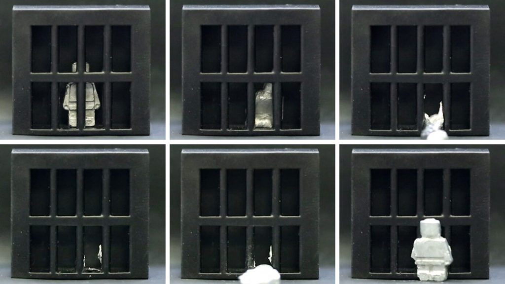
Majidi and colleagues tested their strategy in tiny machines that performed different tasks. In a demonstration straight out of the movie Terminator 2, a toy person escaped a jail cell by melting through the bars and resolidifying in its original form using a mold placed just outside the bars. On the more practical side, one machine removed a small ball from a model human stomach by melting slightly to wrap itself around the foreign object before exiting the organ. But gallium on its own would turn to goo inside a real human body, since the metal is a liquid at body temperature, about 37° C. A few more metals, such as bismuth and tin, would be added to the gallium in biomedical applications to raise the material’s melting point, the authors say. In another demonstration, the material liquefied and rehardened to solder a circuit board. Although this phase-shifting material is a big step in the field, questions remain about its biomedical applications, says biomedical engineer Amir Jafari of the University of North Texas in Denton, who was not involved in the work. One big challenge, he says, is precisely controlling magnetic forces inside the human body that are generated from an external device.
“It’s a compelling tool,” says robotics engineer Nicholas Bira of Harvard University, who was also not involved in the study. But, he adds, scientists who study soft robotics are constantly creating new materials.
“The true innovation to come lies in combining these different innovative materials.”
The Arctic today is a hostile place for most primates. But a series of fossils found since the 1970s suggest that wasn’t always the case.
Dozens of fossilized teeth and jaw bones unearthed in northern Canada belonged to two species of early primates — or at least close relatives of primates — that lived in the Arctic around 52 million years ago, researchers report January 25 in PLOS ONE. These remains are the first primate-like fossils ever discovered in the Arctic and tell of a groundhog-sized animal that may have skittered across trees in a swamp that once existed above the Arctic Circle. The Arctic was significantly warmer during that time. But creatures still had to adapt to extreme conditions such as long winter months without sunlight. These challenges make the presence of primate-like creatures in the Arctic “incredibly surprising,” says coauthor Chris Beard, a paleontologist at the University of Kansas in Lawrence. “No other primate or primate relative has ever been found this far north so far.”
Between frigid temperatures, limited plant growth and months of perpetual darkness, living in the modern Arctic isn’t easy. This is especially true for primates, which evolved from small, tree-dwelling creatures that largely fed on fruit (SN: 6/5/13). To this day, most primates — humans and few other outliers like Japan’s snow monkeys excepted — tend to stick to tropical and subtropical forests, largely found around the equator.
But these forests haven’t always been confined to their present location. During the early Eocene Epoch, which started around 56 million years ago, the planet underwent a period of intense warming that allowed forests and their warm-loving residents to expand northward (SN: 11/3/15).
Scientists know about this early Arctic climate in part because of decades of paleontological work on Ellesmere Island in northern Canada. These digs revealed that the area was once dominated by swamps not unlike those found in the southeastern United States today. This ancient, warm, wet Arctic environment was home to a wide array of heat-loving animals, including giant tapirs and crocodile relatives. For the new study, Beard and his colleagues examined dozens of teeth and jawbone fossils found in the area, concluding that they belong to two species, Ignacius mckennai and Ignacius dawsonae. These two species belonged to a now-extinct genus of small mammals that was widespread across North America during the Eocene. The Arctic variants probably made their way north as the planet warmed, taking advantage of the new habitat opening up near the poles.
Scientists have long debated whether this lineage can be considered true primates or whether they were simply close relatives. Regardless, it’s still “really weird and unexpected” to find primates or their relatives in the area, says Mary Silcox, a vertebrate paleontologist at the University of Toronto Scarborough.
For one thing, Ellesmere Island was already north of the Arctic Circle 52 million years ago. So while conditions may have been warmer and wetter, the swamp was plunged into continuous darkness during the winter months.
Newly arrived Ignacius would have had to adapt to these conditions. Unlike their southern kin, the Arctic Ignacius had unusually strong jaws and teeth suited to eating hard foods, the researchers found. This may have helped these early primates feed on nuts and seeds over the winter, when fruit wasn’t as readily available.
This research can shed light on how animals can adapt to live in extreme conditions. “Ellesmere Island is arguably the best deep time analog for a mild, ice-free Arctic,” says Jaelyn Eberle, a vertebrate paleontologist at the University of Colorado Boulder.
Studying how plants and animals adapted to this remarkable period in Arctic history, Beard says, could offer clues to the Arctic’s future residents.
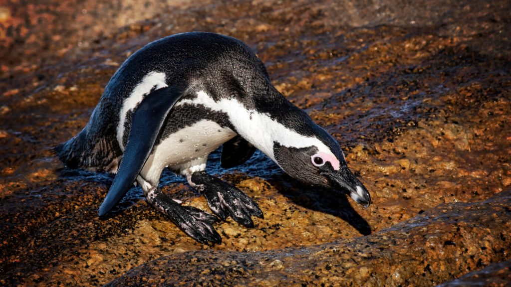
Birds that dive underwater — such as penguins, loons and grebes — may be more likely to go extinct than their nondiving kin, a new study finds.
Many water birds have evolved highly specialized bodies and behaviors that facilitate diving. Now, an analysis of the evolutionary history of more than 700 water bird species shows that once a bird group gains the ability to dive, the change is irreversible. That inflexibility could help explain why diving birds have an elevated extinction rate compared with nondiving birds, researchers report in the Dec. 21 Proceedings of the Royal Society B. “There are substantial morphological adaptations for diving,” says Catherine Sheard, an evolutionary biologist at the University of Bristol in England, who was not involved with the study. For instance, birds that plunge into the water from the air, such as gannets and some pelicans, may have tweaks to the neck muscles and the bones in the chest.
It’s possible that some diving birds are evolving under an evolutionary “ratchet,” where adaptations to exploit a certain food source or habitat unlock some new opportunities, but also encourage ever more specialized evolutionary tailoring. These birds may become trapped in their ways, increasing their risk of extinction. That’s especially true if their habitat rapidly changes in some negative way, possibly because of human-caused climate change (SN: 1/16/20).
Evolutionary biologists Josh Tyler and Jane Younger investigated the evolution of diving in Aequorlitornithes, a collection of 727 water bird species across 11 bird groups. The team divided species into either nondiving birds, or one of three diving types: foot-propelled pursuit (such as loons and grebes), wing-propelled pursuit (like penguins and auks) and the plunge divers.
Diving has evolved at least 14 separate times in the water birds, but there were no instances where diving birds reverted to a nondiving form, the researchers found.
The scientists also explored the link between diving and the development of new species, or their demise, in various bird lineages. Among 236 diving bird species, 75, or 32 percent, were part of lineages that are experiencing 0.02 more species extinctions per million years than the generation of new species. This elevated extinction rate was more common in the wing-propelled and foot-propelled pursuit divers compared with plunge divers. Bird lineages that don’t dive, on the other hand, generated 0.1 more new species per million years than the rate of species dying out.
“The more specialized you become, the more reliant you are on a particular diet, foraging strategy or environment,” says Tyler, of the University of Bath in England. “The range of environments available for foraging is much larger for the nondiving birds than for the specialist divers, and this may play into their ability to adapt and thrive.”
Within diving bird groups, the less specialized, the better. Take penguins, a group that has become the subject of a fair share of conservation concern (SN: 8/1/18). The researchers point out that gentoo penguins (Pygoscelis papua) — which have a broad diet — have larger population sizes than related chinstrap penguins (P. antarcticus) that eat mostly krill, and may actually be as many as four very recently diverged species. The International Union for the Conservation of Nature considers both penguin species to be of “least concern” in terms of imminent extinction risk. But chinstrap numbers are declining in some areas, while gentoo population numbers remain generally stable.
If some diving birds are being trapped in their environments by their own adaptations, that doesn’t bode well for their long-term survival, say Tyler and Younger, who is at the University of Tasmania in Hobart.
According to the IUCN, 156 species, or about one-fifth, of the 727 species of water birds are considered vulnerable, endangered or critically endangered. The researchers calculate that of the 75 diving bird species from lineages with heightened extinction rates, 24 species, or nearly one-third, are already listed as threatened.
We spend so much time making sure wildlife stays away from us, whether that’s setting traps, building fences or putting out poisons. Sure, unwanted guests are annoying. But why do we consider some animals “pests”? It’s all about perspective, says science journalist Bethany Brookshire. “We can put poison out for rats and protest their use as laboratory animals. We can shoot deer in the fall and show their adorable offspring to our children in the spring,” she writes in her new book, Pests: How Humans Create Animal Villains. Brookshire argues that we deem animals “pests” when we fear them (like snakes). Or when they thrive in a niche we unintentionally created for them (think rats in the New York subway). Or when they find a way to live in a habitat now dominated by humans (all those deer in the suburbs). Sometimes we demonize an animal if we feel like it’s threatening our ability to control the landscape (like coyotes that attack our livestock, pets and even children).
Through the lens of science, history, culture, religion, personal anecdotes and a big dose of humor, Brookshire breaks down how our perspective shapes our relationships with our animal neighbors. She also goes into the field — trailing rats, hunting pythons, taming feral cats, tracking drugged-up bears — to see firsthand how pests are treated.
Science News spoke with Brookshire, a former staff writer for Science News for Students (now Science News Explores), about what we can learn from pests and how we can coexist with them. The following conversation has been edited for clarity and brevity.
SN: What inspired you to write this book?
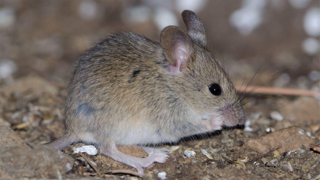
Brookshire: I wrote a news story that was about mice living with humans (SN: 4/19/17). [It was based on a study] showing that we’ve had house mice since we’ve had houses. I love the fact that humans have had these other animals taking advantage of the ecosystems that we create basically since we started living settled life. Every location that has humans has their “rat.” Sometimes that’s a rat, and sometimes it’s a pigeon or a cockatoo or a lizard or a horse. It’s not about what these animals are doing. Animals live in ecosystems that we create, and we hate animals that live too close.
SN: What surprised you during your research?
Brookshire: The reflexiveness of people’s responses [to pests]. People respond emotionally. When you make them pause and think about it, they go, “Oh wow, that doesn’t make any sense. I should not be caught trying to kill a raccoon with a sword.” But in the moment, you’re so wrapped up in the violation of what you see as your personal space.
The other thing is the extent to which our disdain of pests is wrapped up in social justice. A lot of times we see this hatred and disgust for animals that we see as “low class.” High-class people don’t have rats. And that’s really about social justice, about infrastructure and the ability of people to live in clean houses, store their food properly or even have a house at all.
Also, the way we deal with these animals often has vestiges of colonialism, as in the chapter on elephants. [In Kenya, European colonists] made people grow corn and sugarcane, which elephants love. Colonization created national park systems that assumed that humans had no place in wilderness, shoving out Indigenous pastoralists. Colonization created the market for poached ivory. And colonizing people assumed that Indigenous people did not like elephants or know their benefits. We are living with the consequences. Many modern efforts at elephant protection are spearheaded by Western people, and they assume the biggest issue with elephants is poaching and that Indigenous people don’t know what’s best for themselves or the elephants. In fact, human-elephant conflict [which includes elephant crop raids] is the far bigger problem, and Indigenous people have a long history of coexisting with elephants.
SN: In the book, you looked at many different cultures and included Indigenous voices.
Brookshire: It’s important to realize there’s more than one way to look at the world. By learning from other cultures, it helps us understand our biases. It’s only when you get outside of your own beliefs that you realize that’s not just the way things are.
SN: That shows up when you write about the Karni Mata Temple in India, also known as the Temple of Rats. Temple rats are not treated as pests, but a rat in a house would be. Brookshire: That’s the result of context. And you see that in Western cultures all the time. People love squirrels. Well, they’re basically rats with better PR. Then you have people who have pet rats, who would probably scream if a sewer rat ran by.
SN: Are there any animals that you consider a pest?
Brookshire: No. The animal that I’ve probably come away with the most negative impression of is humans. It’s funny because we think we can extinct anything. And I love how these animals have gone: “Oh, poison? That’s cute.” “Oh, a trap? You’re funny.” We’ve tried to use electric fences on elephants [to stop them from eating crops]. And elephants are like, “Guess what? Ivory doesn’t conduct electricity.” Even if they don’t have tusks, elephants just pick up a log [to destroy the fence].
SN: Are you hoping to change people’s minds about pests?
Brookshire: I hope that they will ask why they respond to pests the way they do. Instead of just going, “This animal bothers me,” ask why, and does it make sense. I also hope it opens more curiosity about the animals around us. I learned from Indigenous groups just how much knowledge they have of the animals in their ecosystem. I hope more people learn. A world that you know a lot about is just a better world to live in.
In the battle against the invasive house mouse on islands, scientists are using the rodent’s own genes against it.
With the right tweaks, introducing a few hundred genetically altered mice could drive an island’s invasive mouse population to extinction in about 25 years, researchers report in the Nov. 15 Proceedings of the National Academy of Sciences. The trick is adding the changes to a section of mouse DNA that gets inherited far more often than it should. Scientists have been creating similar extra-inheritable genes — called gene drives — in the lab. The chunks are designed to get passed on to most or all of an animal’s offspring instead of the usual half, and make those offspring infertile in the bargain. Scientists have used gene drives to reduce populations of mosquitoes and fruit flies (SN: 12/17/18).
But mammals are a different story. Scientists have previously synthesized a gene drive that gets passed on in mice about 80 percent of the time (SN: 1/23/19). But the drive isn’t strong enough to stop a population quickly.
Luckily, nature has it handled. A haplotype is a naturally occurring group of genes that gets passed on as a unit during replication. The genome of the house mouse (Mus musculus) has a particular haplotype, called the t haplotype, that gets passed on to offspring more than 95 percent of the time, instead of the typical 50 percent.
This natural gene drive has benefits, says Anna Lindholm, a biologist at the University of Zurich who was not involved in the study. It “evolved naturally and continues to be present in the wild, and we have as yet not found resistance to it in wild populations,” she says. It’s also not found in species besides M. musculus, meaning it probably won’t spread to other noninvasive mice.
Molecular biologist Paul Thomas and his colleagues decided to target the t haplotype with the cut-and-paste molecular tool called CRISPR/Cas9 (SN: 8/24/16). They used CRISPR to insert the gene sequence for the CRISPR tool itself into the t haplotype. When a male mouse carrying the altered t haplotype mates with a female, the inserted genes for the CRISPR tool spring into action. It uses a special genetic guide to target and inactivate the gene for the hormone prolactin — rendering any baby female mice infertile.
The best part is that the natural t haplotype can also sterilize males, says Thomas, of the University of Adelaide in Australia. Males with two copies — homozygous males — won’t reproduce at all.
“If you could get a t to spread through a population, you could get homozygous males being sterile,” he says. “And with the addition of the CRISPR element on top of that, we get homozygous females that are also sterile.” To find out how well the t haplotype mice do on an island where mice are wreaking havoc on biodiversity, the scientists used a computer simulation of an island with 200,000 mice. The team found that adding just 256 mice with the CRISPR-altered t haplotype could successfully drive the mouse population to zero in around 25 years. Even without CRISPR, adding mice with the normal t haplotype could tank the population in about 43 years.
But models aren’t mice. In a final test, Thomas and his colleagues made the model reality. The team altered the t haplotype in a small group of mice in the lab and used genetic tests to show that those mice would pass on their new genetics 95 percent of the time.
“This is a clever idea, to build on the t haplotype natural drive system and use CRISPR, not for spreading the construct, but for damaging genes necessary for female fertility,” Lindholm says. “This is a big advance in the development of new tools to control invasive mouse populations.”
The next step, Thomas says, will be to test the effects in real populations of mice in secure enclosures, to find out if the genetically tweaked t can stop mice from reproducing. The scientists also want to ensure that any engineered mice released into the wild have some safety mechanism in place, so other mice elsewhere remain unaffected.
The final version might target tiny mutations that only occur on one island where the pest population is isolated, Thomas suggests. If the mouse escaped onto the mainland, its altered genes would have no effect on the local mice. The scientists also want to consult with people living in the area, as officials did when genetically modified mosquitoes were released in Florida (SN: 5/14/21).
Finally, he notes, 25 years is a long wait for some endangered island populations. “We would love to see CRISPR work faster,” he says. “It’s still a work in progress.”
Just how hot is your chili pepper? A new chili-shaped device could quickly signal whether adding the pepper to a meal might set your mouth ablaze.
Called the Chilica-pod, the device detects capsaicin, a chemical compound that helps give peppers their sometimes painful kick. In general, the more capsaicin a pepper has, the hotter it tastes. The Chilica-pod is sensitive, capable of detecting extremely low levels of the fiery molecule, researchers report in the Oct. 23 ACS Applied Nano Materials.
The device could someday be used to test cooked meals or fresh peppers, says analytical chemist Warakorn Limbut of Prince of Songkla University in Hat Yai, Thailand. People with a capsaicin allergy could use the gadget to avoid the compound, or farmers could test harvested peppers to better indicate their spiciness, he says. A pepper’s relative spiciness typically is conveyed in Scoville heat units — an imperfect measurement determined by a panel of human taste testers. Other more precise methods for determining spiciness are time-intensive and involve expensive equipment, making the methods unsuitable for a quick answer.
Enter the portable, smartphone-compatible Chilica-pod. Built by Limbut and colleagues, the instrument’s sensor is composed of stacks of graphene sheets. When a drop of a chili pepper and ethanol solution is added to the sensor, the capsaicin from the pepper triggers the movement of electrons among the graphene atoms. The more capsaicin the solution has, the stronger the electrical current through the sheets.
The Chilica-pod registers that electrical activity and, once its “stem” is plugged into a smartphone, sends the information to an app for analysis. The device can detect capsaicin levels as low as 0.37 micromoles per liter of solution, equivalent to the amount in a pepper with no heat, one test showed.
Limbut’s team used the Chilica-pod to individually measure six dried chili peppers from a local market. The peppers’ capsaicin concentrations ranged from 7.5 to 90 micromoles per liter of solution, the team found. When translated to Scoville heat units, that range corresponds to the spice of peppers like serrano or cayenne — mild varieties compared to the blazing hot Carolina reaper, one of the world’s hottest peppers (SN: 4/9/18).
Paul Bosland, a plant geneticist and chili breeder at New Mexico State University in Las Cruces who wasn’t involved in the study, notes that capsaicin is just one of at least 24 related compounds that give peppers heat. “I would hope that [the device] could read them all,” he says.
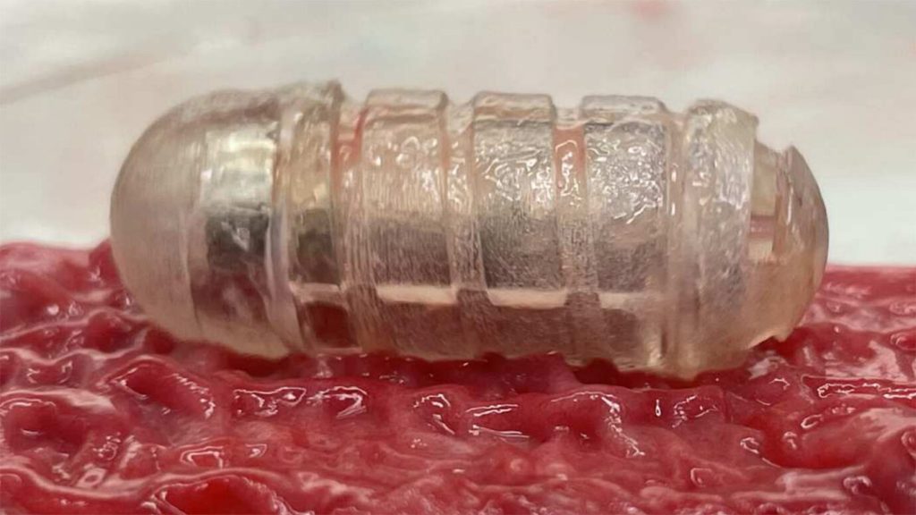
A mucus-wicking robotic pill may offer a new way to deliver meds.
The multivitamin-sized device houses a motor and a cargo hold for drugs, including ones that are typically given via injections or intravenously, such as insulin and some antibiotics. If people could take such drugs orally, they could potentially avoid daily shots or a hospital stay, which would be “a huge game changer,” says MIT biomedical engineer Shriya Srinivasan.
But drugs that enter the body via the mouth face a tough journey. They encounter churning stomach acid, raging digestive enzymes and sticky slicks of mucus in the gut. Intestinal mucus “sort of acts like Jell-O,” Srinivasan says. The goo can trap drug particles, preventing them from entering the bloodstream.
The new device, dubbed RoboCap, whisks away this problem. The pill uses surface grooves, studs and torpedo-inspired fins to scrub away intestinal mucus like a miniature brush whirling inside a bottle. In experiments in pigs, RoboCap tunneled through mucus lining the walls of the small intestine, depositing insulin or the IV antibiotic vancomycin along the way, Srinivasan and colleagues report September 28 in Science Robotics. After churning for about 35 minutes, the pill continued its trip through the gut and eventually out of the body.
RoboCap is the latest pill-like gadget made to be swallowed. In 2019, some of the same researchers who developed RoboCap debuted a different device — one that injects drugs by pricking the inside of the stomach (SN: 2/7/19). That pea-sized injector was not designed to work in the small intestine, where some drugs are most easily absorbed. The RoboCap may also be able to deliver larger drug payloads, Srinivasan says.